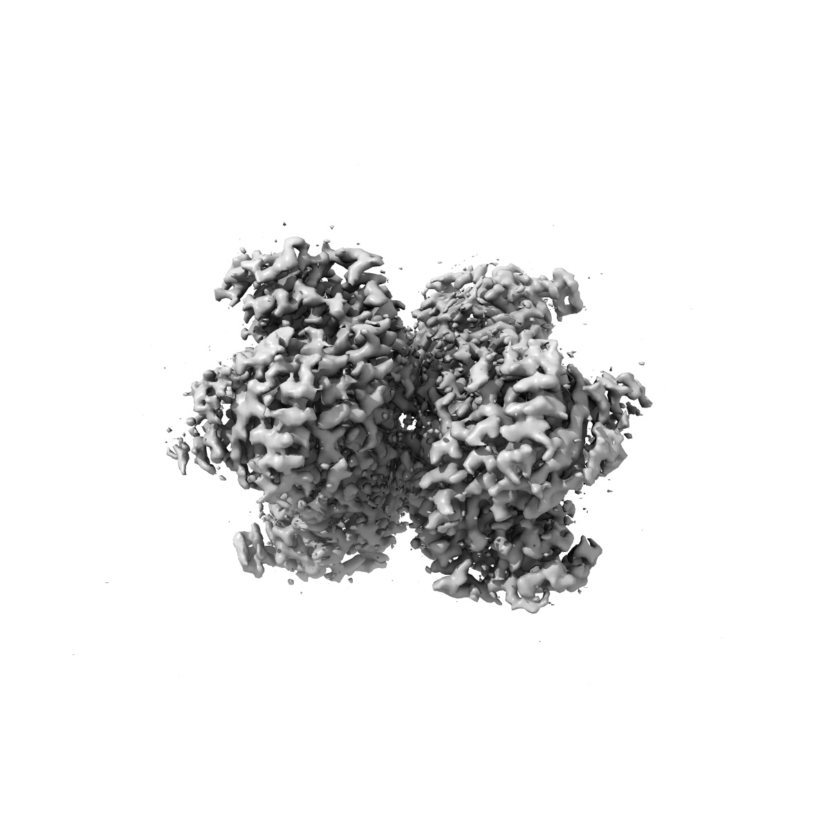
EMD-26442
Structure of G6PD-D200N tetramer bound to NADP+ with no symmetry applied
EMD-26442
Single-particle2.5 Å
 Deposition: 15/03/2022
Map released: 14/09/2022
Last modified: 12/06/2024
Sample Organism:
Homo sapiens
Sample: HUMAN Glucose-6-phosphate 1-dehydrogenase in complex with NADP+
Fitted models: 7uc2 (Avg. Q-score: 0.614)
Deposition Authors: Wei X, Marmorstein R
Sample: HUMAN Glucose-6-phosphate 1-dehydrogenase in complex with NADP+
Fitted models: 7uc2 (Avg. Q-score: 0.614)
Deposition Authors: Wei X, Marmorstein R

Allosteric role of a structural NADP + molecule in glucose-6-phosphate dehydrogenase activity.
Wei X,
Kixmoeller K ,
Baltrusaitis E,
Yang X ,
Marmorstein R
(2022) PNAS , 119 , e2119695119 - e2119695119
,
Baltrusaitis E,
Yang X ,
Marmorstein R
(2022) PNAS , 119 , e2119695119 - e2119695119
Abstract:
Human glucose-6-phosphate dehydrogenase (G6PD) is the main cellular source of NADPH, and thus plays a key role in maintaining reduced glutathione to protect cells from oxidative stress disorders such as hemolytic anemia. G6PD is a multimeric enzyme that uses the cofactors β-D-glucose 6-phosphate (G6P) and "catalytic" NADP+ (NADP+c), as well as a "structural" NADP+ (NADP+s) located ∼25 Å from the active site, to generate NADPH. While X-ray crystallographic and biochemical studies have revealed a role for NADP+s in maintaining the catalytic activity by stabilizing the multimeric G6PD conformation, other potential roles for NADP+s have not been evaluated. Here, we determined the high resolution cryo-electron microscopy structures of human wild-type G6PD in the absence of bound ligands and a catalytic G6PD-D200N mutant bound to NADP+c and NADP+s in the absence or presence of G6P. A comparison of these structures, together with previously reported structures, reveals that the unliganded human G6PD forms a mixture of dimers and tetramers with similar overall folds, and binding of NADP+s induces a structural ordering of a C-terminal extension region and allosterically regulates G6P binding and catalysis. These studies have implications for understanding G6PD deficiencies and for therapy of G6PD-mediated disorders.
Human glucose-6-phosphate dehydrogenase (G6PD) is the main cellular source of NADPH, and thus plays a key role in maintaining reduced glutathione to protect cells from oxidative stress disorders such as hemolytic anemia. G6PD is a multimeric enzyme that uses the cofactors β-D-glucose 6-phosphate (G6P) and "catalytic" NADP+ (NADP+c), as well as a "structural" NADP+ (NADP+s) located ∼25 Å from the active site, to generate NADPH. While X-ray crystallographic and biochemical studies have revealed a role for NADP+s in maintaining the catalytic activity by stabilizing the multimeric G6PD conformation, other potential roles for NADP+s have not been evaluated. Here, we determined the high resolution cryo-electron microscopy structures of human wild-type G6PD in the absence of bound ligands and a catalytic G6PD-D200N mutant bound to NADP+c and NADP+s in the absence or presence of G6P. A comparison of these structures, together with previously reported structures, reveals that the unliganded human G6PD forms a mixture of dimers and tetramers with similar overall folds, and binding of NADP+s induces a structural ordering of a C-terminal extension region and allosterically regulates G6P binding and catalysis. These studies have implications for understanding G6PD deficiencies and for therapy of G6PD-mediated disorders.