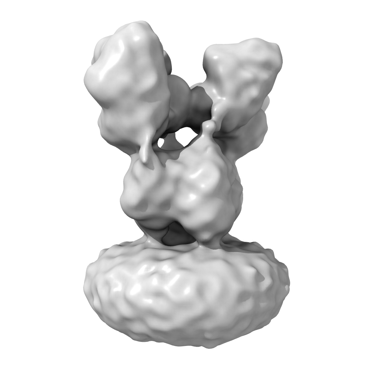
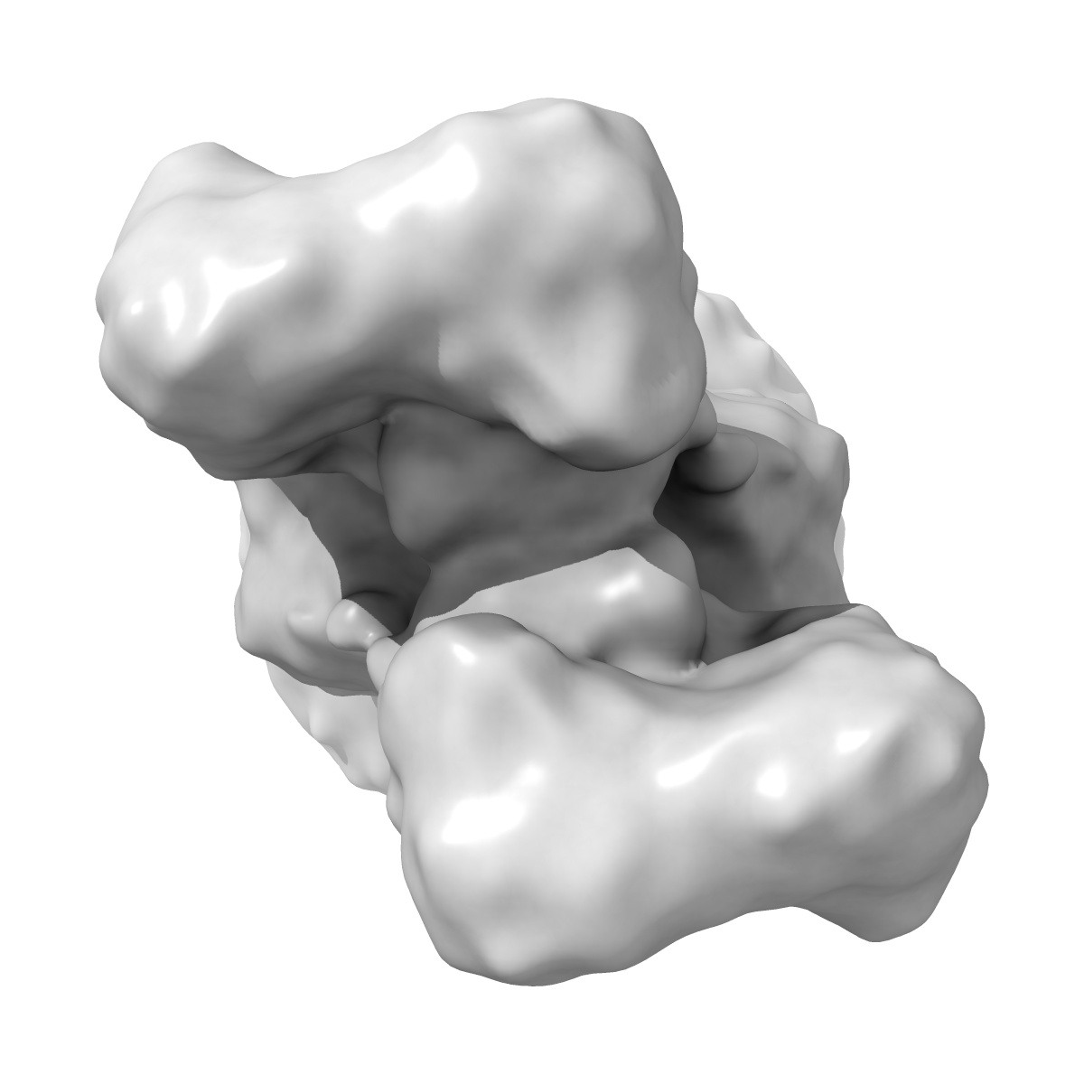
EMD-2684
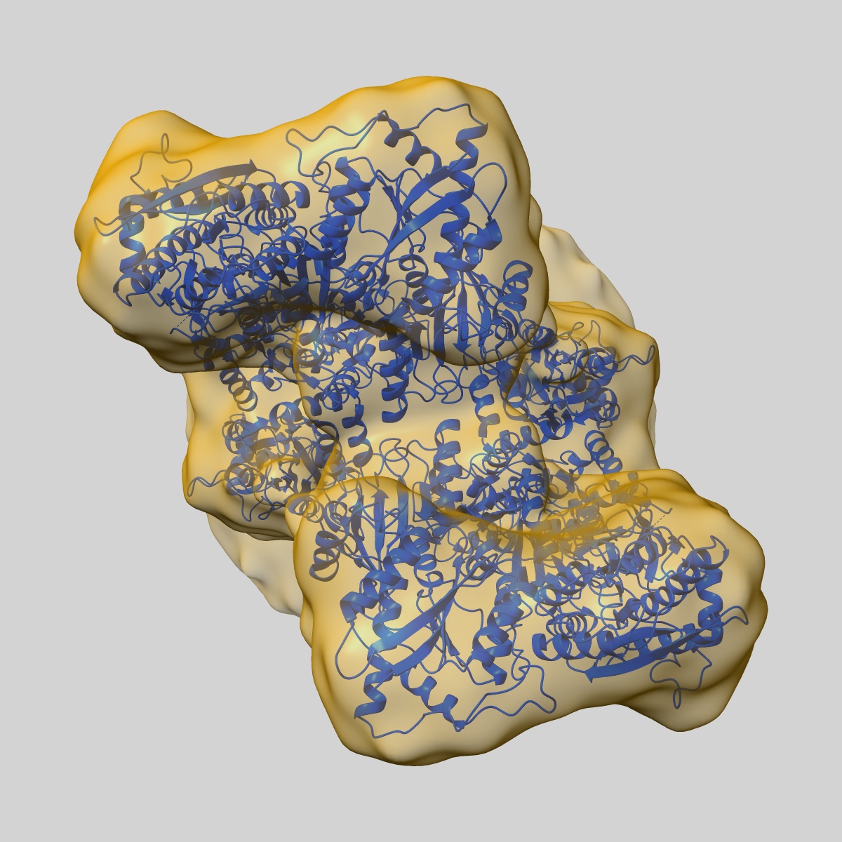
Density map of GluA2em in complex with LY451646 and glutamate
EMD-2684
Single-particle12.8 Å
 Deposition: 20/06/2014
Map released: 13/08/2014
Last modified: 29/10/2014
Sample Organism:
Rattus norvegicus,
synthetic construct
Sample: GluA2em with LY451646 and glutamate
Fitted models: 4uq6 (Avg. Q-score: 0.097)
Deposition Authors: Meyerson JR, Kumar J ,
Chittori S ,
Rao P,
Pierson J,
Bartesaghi A,
Mayer ML,
Subramaniam S
,
Chittori S ,
Rao P,
Pierson J,
Bartesaghi A,
Mayer ML,
Subramaniam S
Sample: GluA2em with LY451646 and glutamate
Fitted models: 4uq6 (Avg. Q-score: 0.097)
Deposition Authors: Meyerson JR, Kumar J
 ,
Chittori S ,
Rao P,
Pierson J,
Bartesaghi A,
Mayer ML,
Subramaniam S
,
Chittori S ,
Rao P,
Pierson J,
Bartesaghi A,
Mayer ML,
Subramaniam S
Structural mechanism of glutamate receptor activation and desensitization
Meyerson JR,
Kumar J ,
Chittori S ,
Rao P,
Pierson J,
Bartesaghi A,
Mayer ML,
Subramaniam S
(2014) Nature , 514 , 328 - 334
,
Chittori S ,
Rao P,
Pierson J,
Bartesaghi A,
Mayer ML,
Subramaniam S
(2014) Nature , 514 , 328 - 334
Abstract:
Ionotropic glutamate receptors are ligand-gated ion channels that mediate excitatory synaptic transmission in the vertebrate brain. To gain a better understanding of how structural changes gate ion flux across the membrane, we trapped rat AMPA (α-amino-3-hydroxy-5-methyl-4-isoxazole propionic acid) and kainate receptor subtypes in their major functional states and analysed the resulting structures using cryo-electron microscopy. We show that transition to the active state involves a 'corkscrew' motion of the receptor assembly, driven by closure of the ligand-binding domain. Desensitization is accompanied by disruption of the amino-terminal domain tetramer in AMPA, but not kainate, receptors with a two-fold to four-fold symmetry transition in the ligand-binding domains in both subtypes. The 7.6 Å structure of a desensitized kainate receptor shows how these changes accommodate channel closing. These findings integrate previous physiological, biochemical and structural analyses of glutamate receptors and provide a molecular explanation for key steps in receptor gating.
Ionotropic glutamate receptors are ligand-gated ion channels that mediate excitatory synaptic transmission in the vertebrate brain. To gain a better understanding of how structural changes gate ion flux across the membrane, we trapped rat AMPA (α-amino-3-hydroxy-5-methyl-4-isoxazole propionic acid) and kainate receptor subtypes in their major functional states and analysed the resulting structures using cryo-electron microscopy. We show that transition to the active state involves a 'corkscrew' motion of the receptor assembly, driven by closure of the ligand-binding domain. Desensitization is accompanied by disruption of the amino-terminal domain tetramer in AMPA, but not kainate, receptors with a two-fold to four-fold symmetry transition in the ligand-binding domains in both subtypes. The 7.6 Å structure of a desensitized kainate receptor shows how these changes accommodate channel closing. These findings integrate previous physiological, biochemical and structural analyses of glutamate receptors and provide a molecular explanation for key steps in receptor gating.