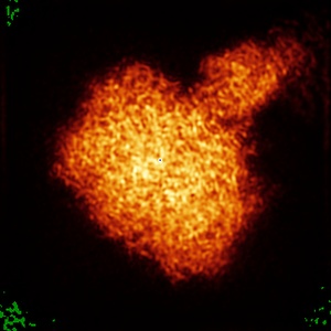
EMD-26949
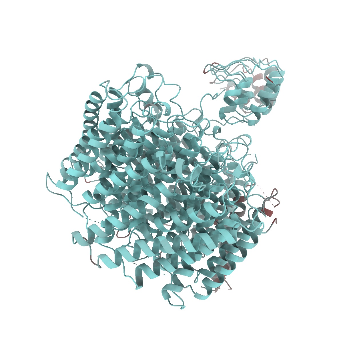
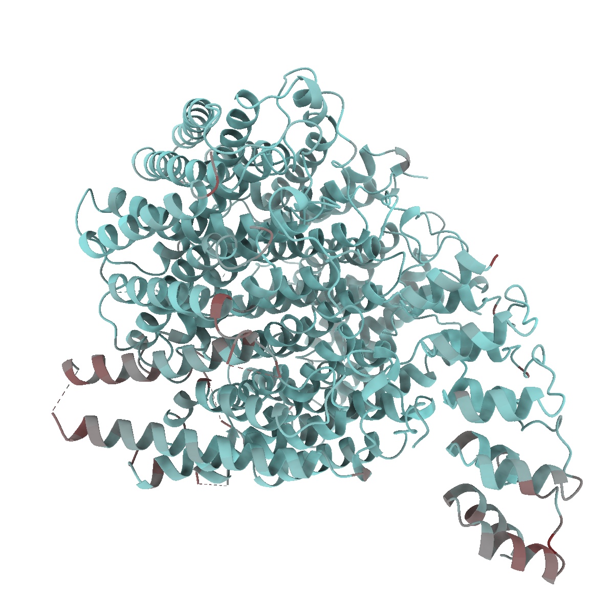
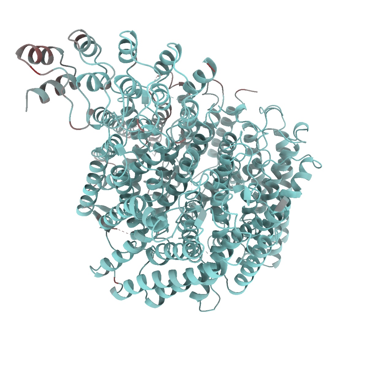
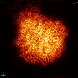
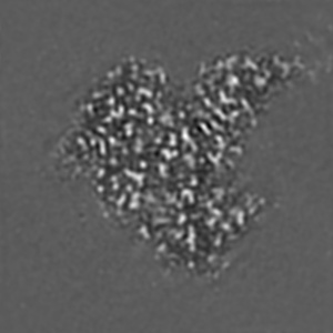
Local refinement of RhAG/CE trimer, class 1 of erythrocyte ankyrin-1 complex
EMD-26949
Single-particle2.5 Å
 Deposition: 10/05/2022
Map released: 20/07/2022
Last modified: 14/02/2024
Sample Organism:
Homo sapiens
Sample: Erythrocyte ankyrin-1 complex
Fitted models: 7v0s (Avg. Q-score: 0.694)
Deposition Authors: Vallese F ,
Kim K ,
Yen LY,
Johnston JD ,
Noble AJ ,
Cali T ,
Clarke OB
,
Kim K ,
Yen LY,
Johnston JD ,
Noble AJ ,
Cali T ,
Clarke OB
Sample: Erythrocyte ankyrin-1 complex
Fitted models: 7v0s (Avg. Q-score: 0.694)
Deposition Authors: Vallese F
 ,
Kim K ,
Yen LY,
Johnston JD ,
Noble AJ ,
Cali T ,
Clarke OB
,
Kim K ,
Yen LY,
Johnston JD ,
Noble AJ ,
Cali T ,
Clarke OB
Architecture of the human erythrocyte ankyrin-1 complex.
Vallese F ,
Kim K ,
Yen LY,
Johnston JD ,
Noble AJ ,
Cali T ,
Clarke OB
(2022) Nat Struct Mol Biol , 29 , 706 - 718
,
Kim K ,
Yen LY,
Johnston JD ,
Noble AJ ,
Cali T ,
Clarke OB
(2022) Nat Struct Mol Biol , 29 , 706 - 718
Abstract:
The stability and shape of the erythrocyte membrane is provided by the ankyrin-1 complex, but how it tethers the spectrin-actin cytoskeleton to the lipid bilayer and the nature of its association with the band 3 anion exchanger and the Rhesus glycoproteins remains unknown. Here we present structures of ankyrin-1 complexes purified from human erythrocytes. We reveal the architecture of a core complex of ankyrin-1, the Rhesus proteins RhAG and RhCE, the band 3 anion exchanger, protein 4.2, glycophorin A and glycophorin B. The distinct T-shaped conformation of membrane-bound ankyrin-1 facilitates recognition of RhCE and, unexpectedly, the water channel aquaporin-1. Together, our results uncover the molecular details of ankyrin-1 association with the erythrocyte membrane, and illustrate the mechanism of ankyrin-mediated membrane protein clustering.
The stability and shape of the erythrocyte membrane is provided by the ankyrin-1 complex, but how it tethers the spectrin-actin cytoskeleton to the lipid bilayer and the nature of its association with the band 3 anion exchanger and the Rhesus glycoproteins remains unknown. Here we present structures of ankyrin-1 complexes purified from human erythrocytes. We reveal the architecture of a core complex of ankyrin-1, the Rhesus proteins RhAG and RhCE, the band 3 anion exchanger, protein 4.2, glycophorin A and glycophorin B. The distinct T-shaped conformation of membrane-bound ankyrin-1 facilitates recognition of RhCE and, unexpectedly, the water channel aquaporin-1. Together, our results uncover the molecular details of ankyrin-1 association with the erythrocyte membrane, and illustrate the mechanism of ankyrin-mediated membrane protein clustering.