EMD-2706
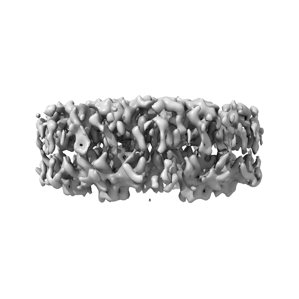
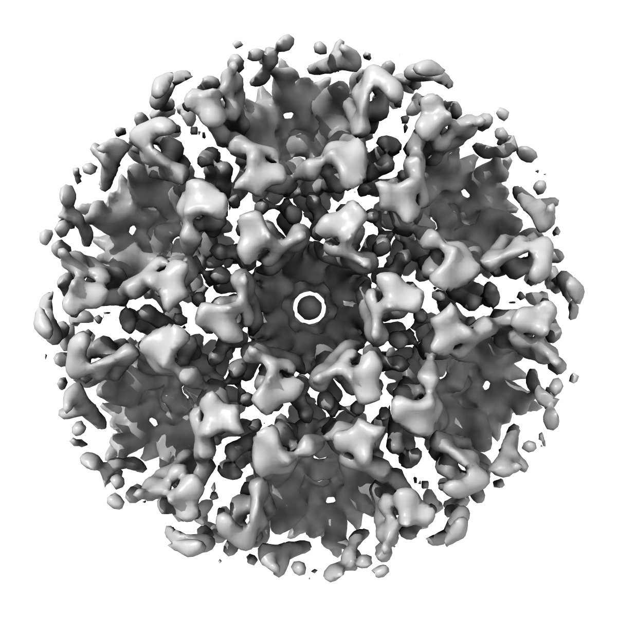
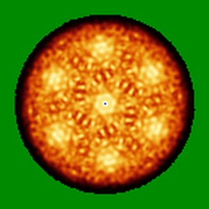
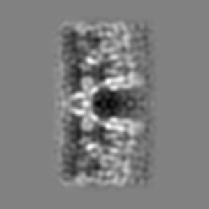
The structure of the immature HIV-1 capsid in intact virus particles at sub-nm resolution
EMD-2706
Subtomogram averaging8.8 Å
 Deposition: 10/07/2014
Map released: 05/11/2014
Last modified: 21/01/2015
Sample Organism:
Human immunodeficiency virus 1
Sample: intact HIV-1 particles treated with the protease inhibitor Amprenavir
Fitted models: 4usn (Avg. Q-score: 0.314)
Deposition Authors: Schur FKM ,
Hagen WJH ,
Rumlova M ,
Ruml T ,
Mueller B ,
Kraeusslich H-G ,
Briggs JAG
,
Hagen WJH ,
Rumlova M ,
Ruml T ,
Mueller B ,
Kraeusslich H-G ,
Briggs JAG
Sample: intact HIV-1 particles treated with the protease inhibitor Amprenavir
Fitted models: 4usn (Avg. Q-score: 0.314)
Deposition Authors: Schur FKM
 ,
Hagen WJH ,
Rumlova M ,
Ruml T ,
Mueller B ,
Kraeusslich H-G ,
Briggs JAG
,
Hagen WJH ,
Rumlova M ,
Ruml T ,
Mueller B ,
Kraeusslich H-G ,
Briggs JAG
Structure of the immature HIV-1 capsid in intact virus particles at 8.8A resolution
Schur FKM ,
Hagen WJH ,
Rumlova M ,
Ruml T ,
Mueller B ,
Kraeusslich H-G ,
Briggs JAG
(2015) Nature , 517 , 505 - 508
,
Hagen WJH ,
Rumlova M ,
Ruml T ,
Mueller B ,
Kraeusslich H-G ,
Briggs JAG
(2015) Nature , 517 , 505 - 508
Abstract:
Human immunodeficiency virus type 1 (HIV-1) assembly proceeds in two stages. First, the 55 kilodalton viral Gag polyprotein assembles into a hexameric protein lattice at the plasma membrane of the infected cell, inducing budding and release of an immature particle. Second, Gag is cleaved by the viral protease, leading to internal rearrangement of the virus into the mature, infectious form. Immature and mature HIV-1 particles are heterogeneous in size and morphology, preventing high-resolution analysis of their protein arrangement in situ by conventional structural biology methods. Here we apply cryo-electron tomography and sub-tomogram averaging methods to resolve the structure of the capsid lattice within intact immature HIV-1 particles at subnanometre resolution, allowing unambiguous positioning of all α-helices. The resulting model reveals tertiary and quaternary structural interactions that mediate HIV-1 assembly. Strikingly, these interactions differ from those predicted by the current model based on in vitro-assembled arrays of Gag-derived proteins from Mason-Pfizer monkey virus. To validate this difference, we solve the structure of the capsid lattice within intact immature Mason-Pfizer monkey virus particles. Comparison with the immature HIV-1 structure reveals that retroviral capsid proteins, while having conserved tertiary structures, adopt different quaternary arrangements during virus assembly. The approach demonstrated here should be applicable to determine structures of other proteins at subnanometre resolution within heterogeneous environments.
Human immunodeficiency virus type 1 (HIV-1) assembly proceeds in two stages. First, the 55 kilodalton viral Gag polyprotein assembles into a hexameric protein lattice at the plasma membrane of the infected cell, inducing budding and release of an immature particle. Second, Gag is cleaved by the viral protease, leading to internal rearrangement of the virus into the mature, infectious form. Immature and mature HIV-1 particles are heterogeneous in size and morphology, preventing high-resolution analysis of their protein arrangement in situ by conventional structural biology methods. Here we apply cryo-electron tomography and sub-tomogram averaging methods to resolve the structure of the capsid lattice within intact immature HIV-1 particles at subnanometre resolution, allowing unambiguous positioning of all α-helices. The resulting model reveals tertiary and quaternary structural interactions that mediate HIV-1 assembly. Strikingly, these interactions differ from those predicted by the current model based on in vitro-assembled arrays of Gag-derived proteins from Mason-Pfizer monkey virus. To validate this difference, we solve the structure of the capsid lattice within intact immature Mason-Pfizer monkey virus particles. Comparison with the immature HIV-1 structure reveals that retroviral capsid proteins, while having conserved tertiary structures, adopt different quaternary arrangements during virus assembly. The approach demonstrated here should be applicable to determine structures of other proteins at subnanometre resolution within heterogeneous environments.