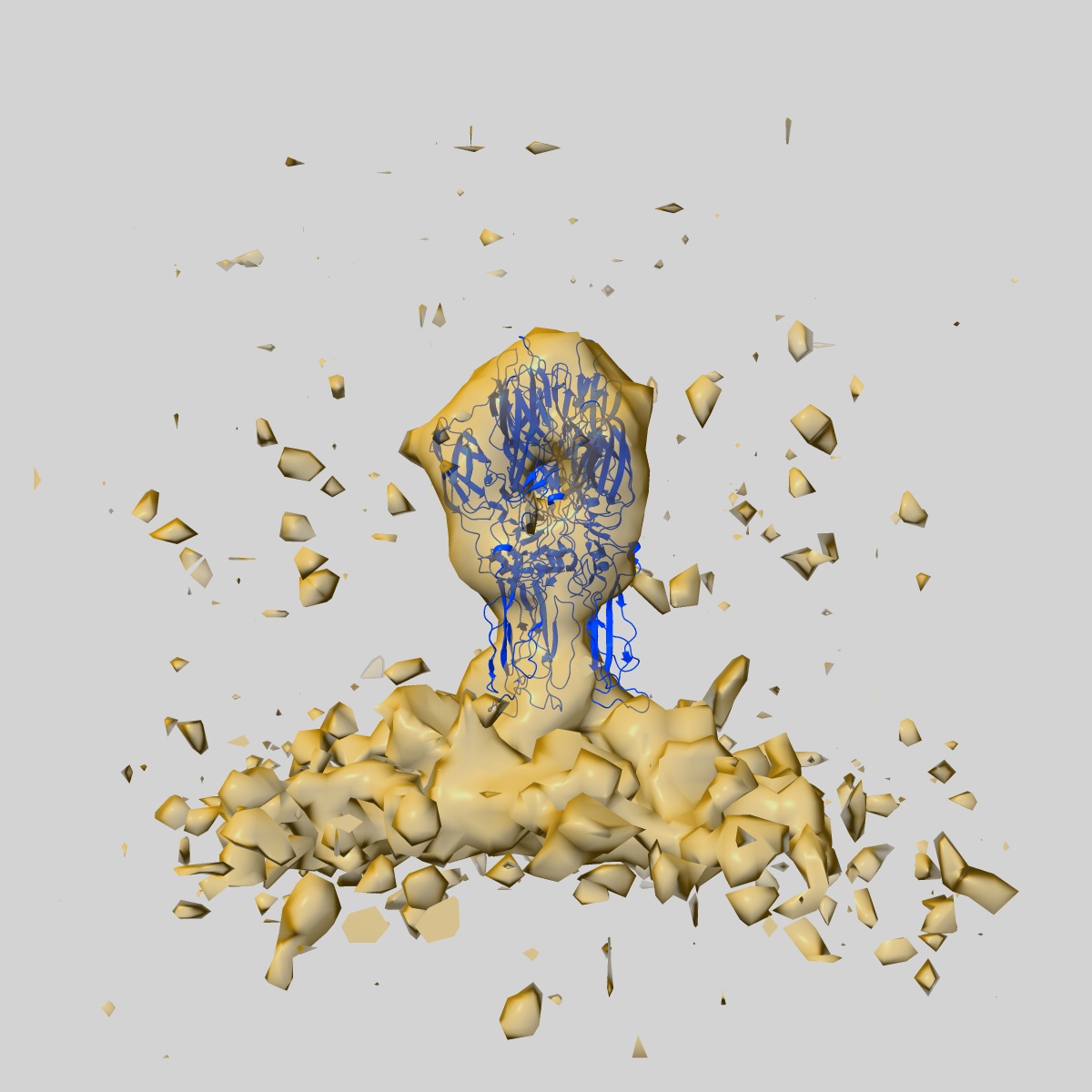
EMD-27248
Sub-tomogram averaged map of full-length post-fusion CHIKV E1 glycoprotein trimer
EMD-27248
Subtomogram averaging27.2 Å
 Deposition: 08/06/2022
Map released: 31/08/2022
Last modified: 23/10/2024
Sample Organism:
Chikungunya virus strain S27-African prototype
Sample: Sub-tomogram averaged map of post-fusion E1 glycoprotein trimer from Chikungunya virus
Fitted models: 8d87 (Avg. Q-score: 0.029)
Deposition Authors: Mangala Prasad V, Lee KK
Sample: Sub-tomogram averaged map of post-fusion E1 glycoprotein trimer from Chikungunya virus
Fitted models: 8d87 (Avg. Q-score: 0.029)
Deposition Authors: Mangala Prasad V, Lee KK

Visualization of conformational changes and membrane remodeling leading to genome delivery by viral class-II fusion machinery.
Abstract:
Chikungunya virus (CHIKV) is a human pathogen that delivers its genome to the host cell cytoplasm through endocytic low pH-activated membrane fusion mediated by class-II fusion proteins. Though structures of prefusion, icosahedral CHIKV are available, structural characterization of virion interaction with membranes has been limited. Here, we have used cryo-electron tomography to visualize CHIKV's complete membrane fusion pathway, identifying key intermediary glycoprotein conformations coupled to membrane remodeling events. Using sub-tomogram averaging, we elucidate features of the low pH-exposed virion, nucleocapsid and full-length E1-glycoprotein's post-fusion structure. Contrary to class-I fusion systems, CHIKV achieves membrane apposition by protrusion of extended E1-glycoprotein homotrimers into the target membrane. The fusion process also features a large hemifusion diaphragm that transitions to a wide pore for intact nucleocapsid delivery. Our analyses provide comprehensive ultrastructural insights into the class-II virus fusion system function and direct mechanistic characterization of the fundamental process of protein-mediated membrane fusion.
Chikungunya virus (CHIKV) is a human pathogen that delivers its genome to the host cell cytoplasm through endocytic low pH-activated membrane fusion mediated by class-II fusion proteins. Though structures of prefusion, icosahedral CHIKV are available, structural characterization of virion interaction with membranes has been limited. Here, we have used cryo-electron tomography to visualize CHIKV's complete membrane fusion pathway, identifying key intermediary glycoprotein conformations coupled to membrane remodeling events. Using sub-tomogram averaging, we elucidate features of the low pH-exposed virion, nucleocapsid and full-length E1-glycoprotein's post-fusion structure. Contrary to class-I fusion systems, CHIKV achieves membrane apposition by protrusion of extended E1-glycoprotein homotrimers into the target membrane. The fusion process also features a large hemifusion diaphragm that transitions to a wide pore for intact nucleocapsid delivery. Our analyses provide comprehensive ultrastructural insights into the class-II virus fusion system function and direct mechanistic characterization of the fundamental process of protein-mediated membrane fusion.