EMD-2804
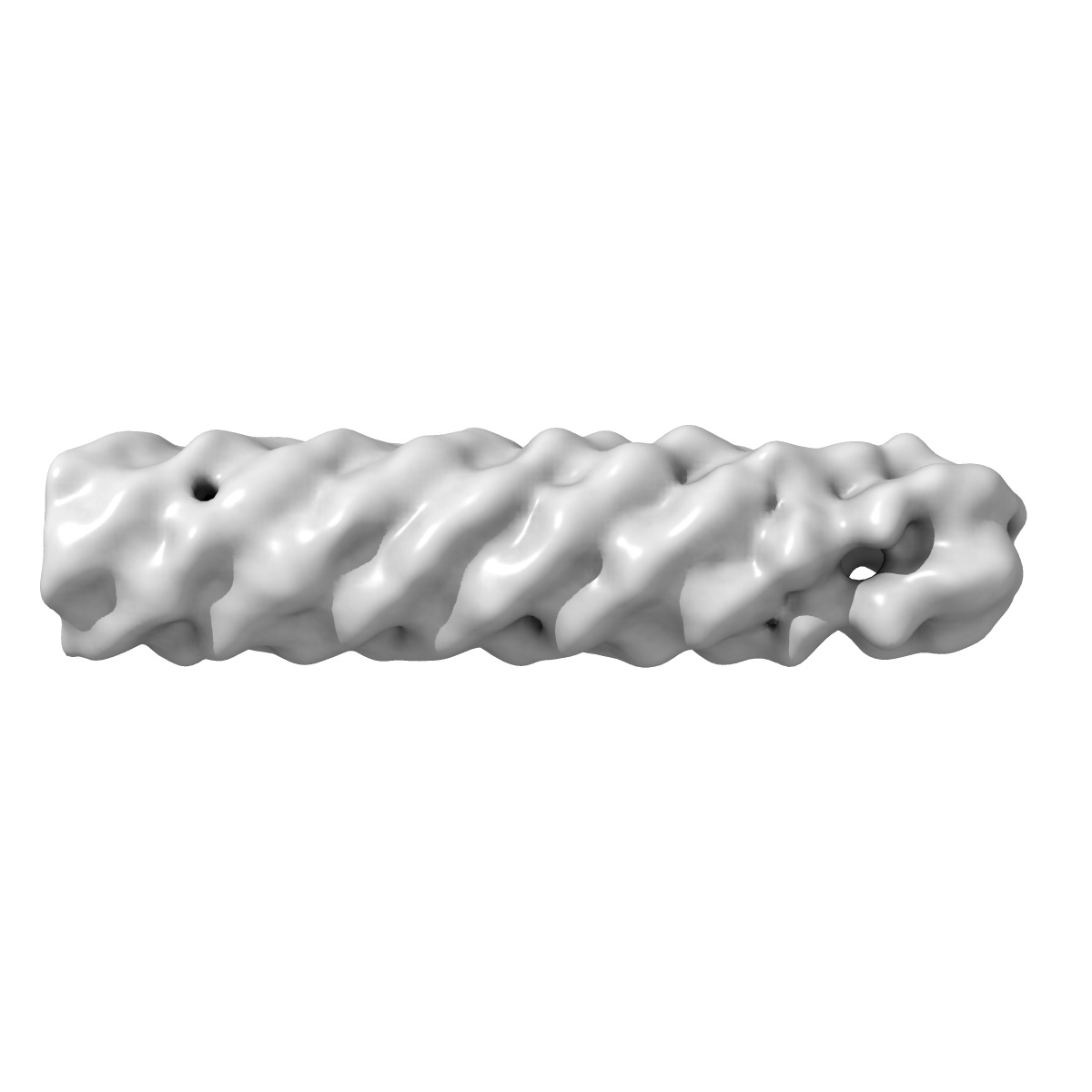
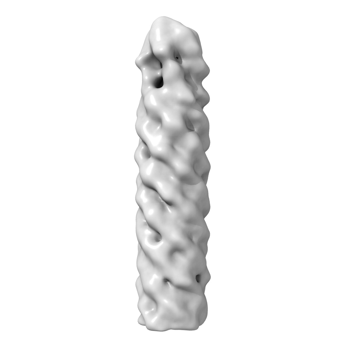
Negative stain electron microscopy reconstruction of the tip complex from the type III secretion system of Shigella flexneri with MxiH mutation Q51A
EMD-2804
Single-particle21.0 Å
 Deposition: 17/10/2014
Map released: 05/11/2014
Last modified: 11/03/2015
Sample Organism:
Shigella flexneri
Sample: Conformation of the tip complex from the type III secretion system of Shigella flexneri bound to the needle with MxiH mutation Q51A
Deposition Authors: Cheung M, Shen D, Makino F, Kato T, Roehrich D, Martinez-Argudo I ,
Walker ML,
Murillo I,
Liu X,
Pain M,
Brown J,
Frazer G,
Mantell J ,
Mina P,
Todd T,
Sessions RB ,
Namba K,
Blocker AJ
,
Walker ML,
Murillo I,
Liu X,
Pain M,
Brown J,
Frazer G,
Mantell J ,
Mina P,
Todd T,
Sessions RB ,
Namba K,
Blocker AJ
Sample: Conformation of the tip complex from the type III secretion system of Shigella flexneri bound to the needle with MxiH mutation Q51A
Deposition Authors: Cheung M, Shen D, Makino F, Kato T, Roehrich D, Martinez-Argudo I
 ,
Walker ML,
Murillo I,
Liu X,
Pain M,
Brown J,
Frazer G,
Mantell J ,
Mina P,
Todd T,
Sessions RB ,
Namba K,
Blocker AJ
,
Walker ML,
Murillo I,
Liu X,
Pain M,
Brown J,
Frazer G,
Mantell J ,
Mina P,
Todd T,
Sessions RB ,
Namba K,
Blocker AJ
Three-dimensional electron microscopy reconstruction and cysteine-mediated crosslinking provide a model of the type III secretion system needle tip complex.
Cheung M,
Shen DK,
Makino F,
Kato T,
Roehrich AD,
Martinez-Argudo I ,
Walker ML,
Murillo I,
Liu X,
Pain M,
Brown J,
Frazer G,
Mantell J ,
Mina P,
Todd T,
Sessions RB ,
Namba K,
Blocker AJ
(2015) MOL.MICROBIOL. , 95 , 31 - 50
,
Walker ML,
Murillo I,
Liu X,
Pain M,
Brown J,
Frazer G,
Mantell J ,
Mina P,
Todd T,
Sessions RB ,
Namba K,
Blocker AJ
(2015) MOL.MICROBIOL. , 95 , 31 - 50
Abstract:
Type III secretion systems are found in many Gram-negative bacteria. They are activated by contact with eukaryotic cells and inject virulence proteins inside them. Host cell detection requires a protein complex located at the tip of the device's external injection needle. The Shigella tip complex (TC) is composed of IpaD, a hydrophilic protein, and IpaB, a hydrophobic protein, which later forms part of the injection pore in the host membrane. Here we used labelling and crosslinking methods to show that TCs from a ΔipaB strain contain five IpaD subunits while the TCs from wild-type can also contain one IpaB and four IpaD subunits. Electron microscopy followed by single particle and helical image analysis was used to reconstruct three-dimensional images of TCs at ∼ 20 Å resolution. Docking of an IpaD crystal structure, constrained by the crosslinks observed, reveals that TC organisation is different from that of all previously proposed models. Our findings suggest new mechanisms for TC assembly and function. The TC is the only site within these secretion systems targeted by disease-protecting antibodies. By suggesting how these act, our work will allow improvement of prophylactic and therapeutic strategies.
Type III secretion systems are found in many Gram-negative bacteria. They are activated by contact with eukaryotic cells and inject virulence proteins inside them. Host cell detection requires a protein complex located at the tip of the device's external injection needle. The Shigella tip complex (TC) is composed of IpaD, a hydrophilic protein, and IpaB, a hydrophobic protein, which later forms part of the injection pore in the host membrane. Here we used labelling and crosslinking methods to show that TCs from a ΔipaB strain contain five IpaD subunits while the TCs from wild-type can also contain one IpaB and four IpaD subunits. Electron microscopy followed by single particle and helical image analysis was used to reconstruct three-dimensional images of TCs at ∼ 20 Å resolution. Docking of an IpaD crystal structure, constrained by the crosslinks observed, reveals that TC organisation is different from that of all previously proposed models. Our findings suggest new mechanisms for TC assembly and function. The TC is the only site within these secretion systems targeted by disease-protecting antibodies. By suggesting how these act, our work will allow improvement of prophylactic and therapeutic strategies.
Secondary citations:
- Veenendaal AK, Hodgkinson JL, Schwarzer L, Stabat D, Zenk SF & Blocker AJ. (2007) The type III secretion system needle tip complex mediates host cell sensing and translocon insertion. MOL.MICROBIOL., 63, 1719 - 1730
- Blocker AJ, Deane JE, Veenendaal AK, Roversi P, Hodgkinson JL, Johnson S & Lea SM. (2008) What's the point of the type III secretion system needle?. PNAS, 105, 6507 - 6513