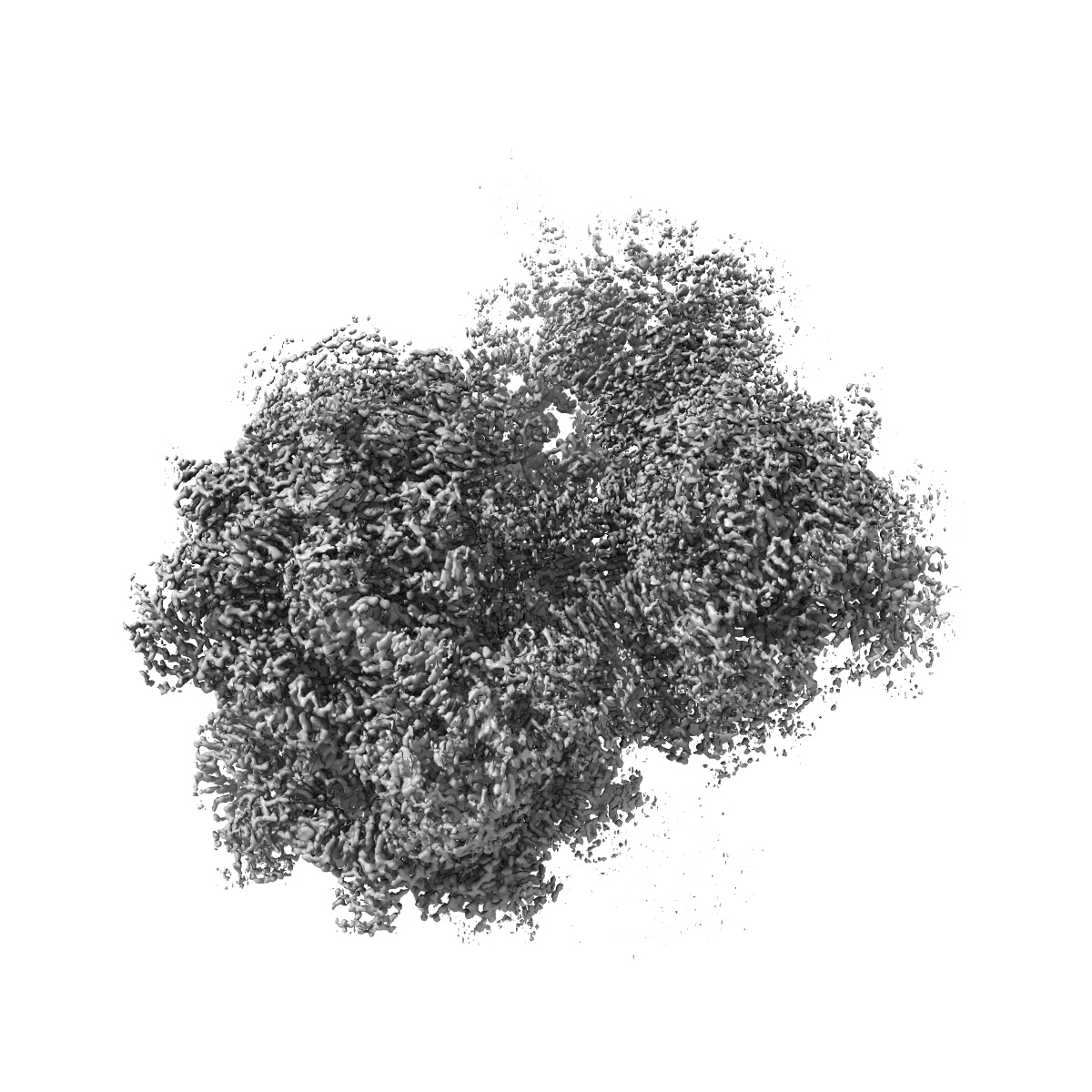
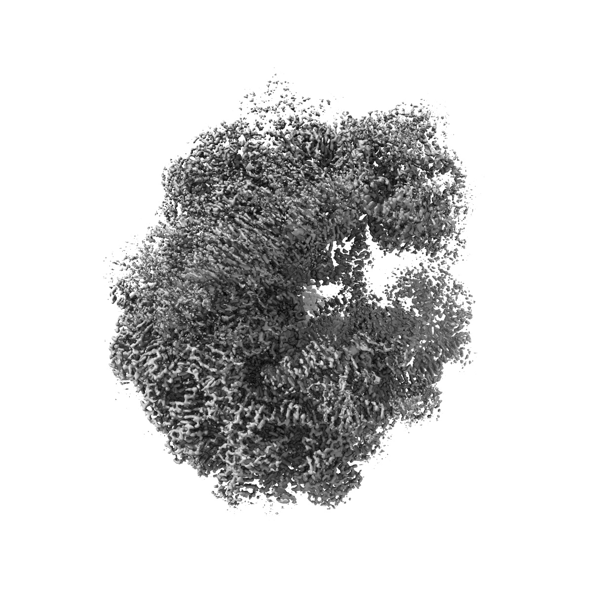
EMD-28255
70S map for: "Atomistic simulations of the E. coli ribosome provide selection criteria for translationally active substrates"
EMD-28255
Single-particle2.2 Å
 Deposition: 28/09/2022
Map released: 31/05/2023
Last modified: 19/07/2023
Sample Organism:
Escherichia coli
Sample: 70S ribosome complex with mRNA, A- and P-site Met-NH-tRNAs
Deposition Authors: Watson ZL ,
Cate JHD
,
Cate JHD
Sample: 70S ribosome complex with mRNA, A- and P-site Met-NH-tRNAs
Deposition Authors: Watson ZL
 ,
Cate JHD
,
Cate JHD
Atomistic simulations of the Escherichia coli ribosome provide selection criteria for translationally active substrates.
Watson ZL ,
Knudson IJ,
Ward FR ,
Miller SJ ,
Cate JHD ,
Schepartz A ,
Abramyan AM
(2023) Nat Chem , 15 , 913 - 921
,
Knudson IJ,
Ward FR ,
Miller SJ ,
Cate JHD ,
Schepartz A ,
Abramyan AM
(2023) Nat Chem , 15 , 913 - 921
Abstract:
As genetic code expansion advances beyond L-α-amino acids to backbone modifications and new polymerization chemistries, delineating what substrates the ribosome can accommodate remains a challenge. The Escherichia coli ribosome tolerates non-L-α-amino acids in vitro, but few structural insights that explain how are available, and the boundary conditions for efficient bond formation are so far unknown. Here we determine a high-resolution cryogenic electron microscopy structure of the E. coli ribosome containing α-amino acid monomers and use metadynamics simulations to define energy surface minima and understand incorporation efficiencies. Reactive monomers across diverse structural classes favour a conformational space where the aminoacyl-tRNA nucleophile is <4 Å from the peptidyl-tRNA carbonyl with a Bürgi-Dunitz angle of 76-115°. Monomers with free energy minima that fall outside this conformational space do not react efficiently. This insight should accelerate the in vivo and in vitro ribosomal synthesis of sequence-defined, non-peptide heterooligomers.
As genetic code expansion advances beyond L-α-amino acids to backbone modifications and new polymerization chemistries, delineating what substrates the ribosome can accommodate remains a challenge. The Escherichia coli ribosome tolerates non-L-α-amino acids in vitro, but few structural insights that explain how are available, and the boundary conditions for efficient bond formation are so far unknown. Here we determine a high-resolution cryogenic electron microscopy structure of the E. coli ribosome containing α-amino acid monomers and use metadynamics simulations to define energy surface minima and understand incorporation efficiencies. Reactive monomers across diverse structural classes favour a conformational space where the aminoacyl-tRNA nucleophile is <4 Å from the peptidyl-tRNA carbonyl with a Bürgi-Dunitz angle of 76-115°. Monomers with free energy minima that fall outside this conformational space do not react efficiently. This insight should accelerate the in vivo and in vitro ribosomal synthesis of sequence-defined, non-peptide heterooligomers.