EMD-28375
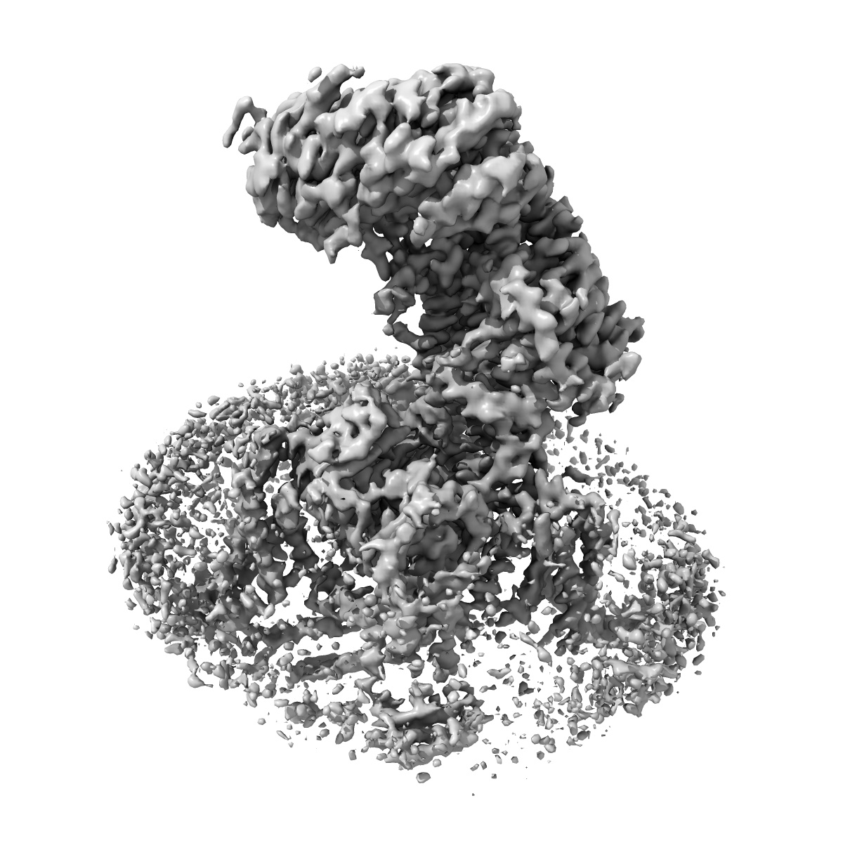
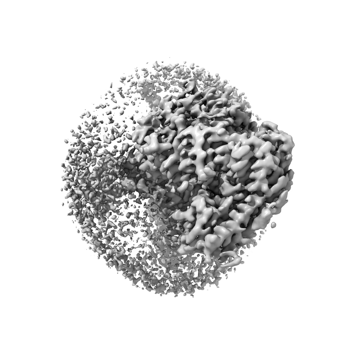
Structure of the human L-type voltage-gated calcium channel Cav1.2 complexed with L-leucine (CABAD Map 2)
EMD-28375
Single-particle3.3 Å
 Deposition: 03/10/2022
Map released: 24/05/2023
Last modified: 23/10/2024
Sample Organism:
Oryctolagus cuniculus,
Homo sapiens
Sample: Ternary complex of human CaV alpha1C with rabbit CaV alpha2delta-1 and rabbit CaV beta3
Fitted models: 8eog (Avg. Q-score: 0.412)
Deposition Authors: Chen Z ,
Mondal A ,
Abderemane-Ali F ,
Minor DL
,
Mondal A ,
Abderemane-Ali F ,
Minor DL
Sample: Ternary complex of human CaV alpha1C with rabbit CaV alpha2delta-1 and rabbit CaV beta3
Fitted models: 8eog (Avg. Q-score: 0.412)
Deposition Authors: Chen Z
 ,
Mondal A ,
Abderemane-Ali F ,
Minor DL
,
Mondal A ,
Abderemane-Ali F ,
Minor DL
EMC chaperone-Ca V structure reveals an ion channel assembly intermediate.
Chen Z ,
Mondal A ,
Abderemane-Ali F ,
Jang S,
Niranjan S,
Montano JL,
Zaro BW ,
Minor Jr DL
(2023) Nature , 619 , 410 - 419
,
Mondal A ,
Abderemane-Ali F ,
Jang S,
Niranjan S,
Montano JL,
Zaro BW ,
Minor Jr DL
(2023) Nature , 619 , 410 - 419
Abstract:
Voltage-gated ion channels (VGICs) comprise multiple structural units, the assembly of which is required for function1,2. Structural understanding of how VGIC subunits assemble and whether chaperone proteins are required is lacking. High-voltage-activated calcium channels (CaVs)3,4 are paradigmatic multisubunit VGICs whose function and trafficking are powerfully shaped by interactions between pore-forming CaV1 or CaV2 CaVα1 (ref. 3), and the auxiliary CaVβ5 and CaVα2δ subunits6,7. Here we present cryo-electron microscopy structures of human brain and cardiac CaV1.2 bound with CaVβ3 to a chaperone-the endoplasmic reticulum membrane protein complex (EMC)8,9-and of the assembled CaV1.2-CaVβ3-CaVα2δ-1 channel. These structures provide a view of an EMC-client complex and define EMC sites-the transmembrane (TM) and cytoplasmic (Cyto) docks; interaction between these sites and the client channel causes partial extraction of a pore subunit and splays open the CaVα2δ-interaction site. The structures identify the CaVα2δ-binding site for gabapentinoid anti-pain and anti-anxiety drugs6, show that EMC and CaVα2δ interactions with the channel are mutually exclusive, and indicate that EMC-to-CaVα2δ hand-off involves a divalent ion-dependent step and CaV1.2 element ordering. Disruption of the EMC-CaV complex compromises CaV function, suggesting that the EMC functions as a channel holdase that facilitates channel assembly. Together, the structures reveal a CaV assembly intermediate and EMC client-binding sites that could have wide-ranging implications for the biogenesis of VGICs and other membrane proteins.
Voltage-gated ion channels (VGICs) comprise multiple structural units, the assembly of which is required for function1,2. Structural understanding of how VGIC subunits assemble and whether chaperone proteins are required is lacking. High-voltage-activated calcium channels (CaVs)3,4 are paradigmatic multisubunit VGICs whose function and trafficking are powerfully shaped by interactions between pore-forming CaV1 or CaV2 CaVα1 (ref. 3), and the auxiliary CaVβ5 and CaVα2δ subunits6,7. Here we present cryo-electron microscopy structures of human brain and cardiac CaV1.2 bound with CaVβ3 to a chaperone-the endoplasmic reticulum membrane protein complex (EMC)8,9-and of the assembled CaV1.2-CaVβ3-CaVα2δ-1 channel. These structures provide a view of an EMC-client complex and define EMC sites-the transmembrane (TM) and cytoplasmic (Cyto) docks; interaction between these sites and the client channel causes partial extraction of a pore subunit and splays open the CaVα2δ-interaction site. The structures identify the CaVα2δ-binding site for gabapentinoid anti-pain and anti-anxiety drugs6, show that EMC and CaVα2δ interactions with the channel are mutually exclusive, and indicate that EMC-to-CaVα2δ hand-off involves a divalent ion-dependent step and CaV1.2 element ordering. Disruption of the EMC-CaV complex compromises CaV function, suggesting that the EMC functions as a channel holdase that facilitates channel assembly. Together, the structures reveal a CaV assembly intermediate and EMC client-binding sites that could have wide-ranging implications for the biogenesis of VGICs and other membrane proteins.