EMD-28713
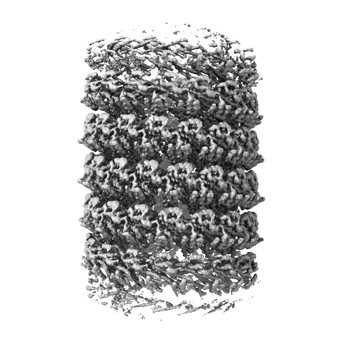
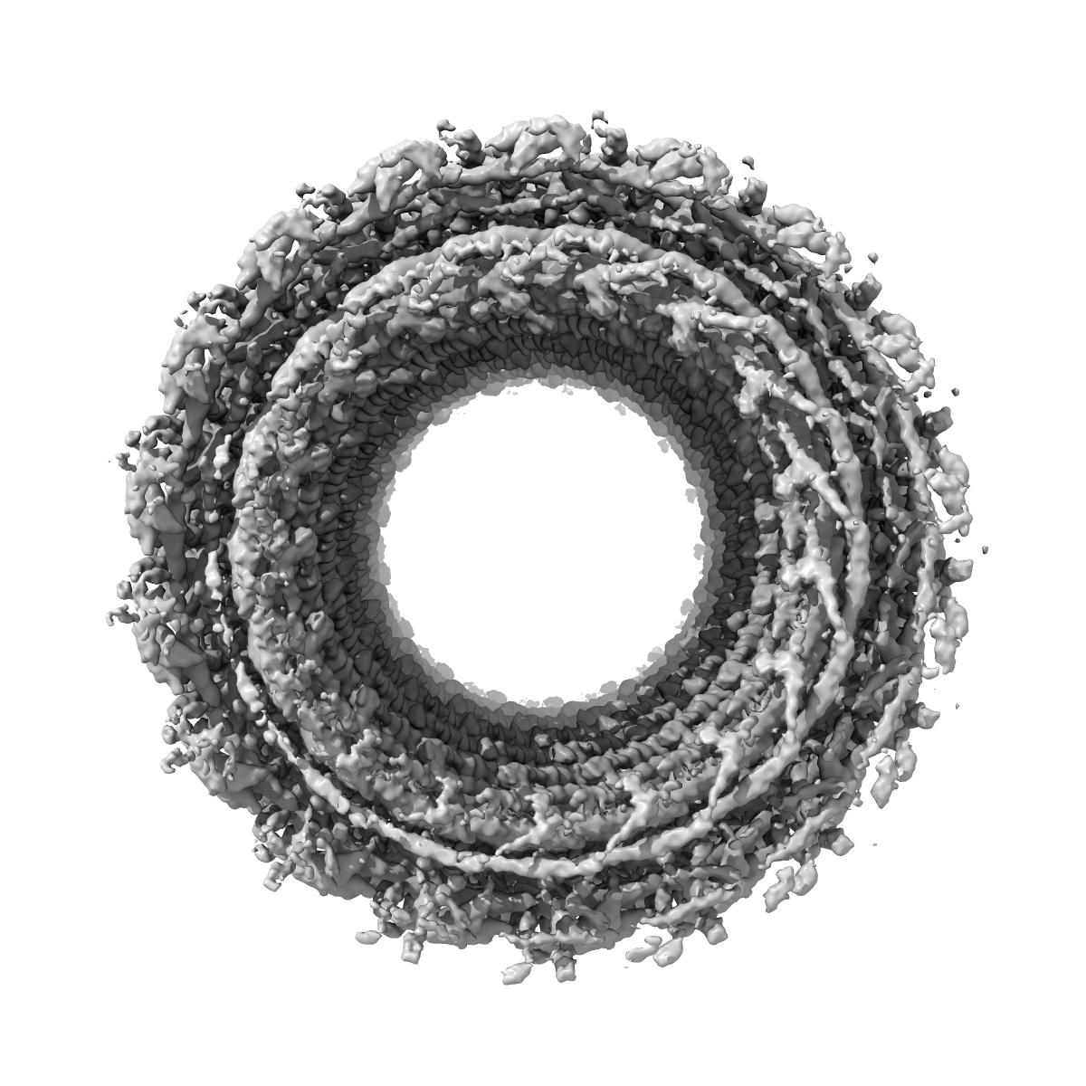
CryoEM reconstruction of membrane-bound, left-handed CHMP1B(F9A/F13A)+IST1 filament with brominated SDPC probe
EMD-28713
Helical reconstruction2.9 Å
 Deposition: 28/10/2022
Map released: 11/01/2023
Last modified: 17/01/2024
Sample Organism:
Homo sapiens
Sample: Helical copolymer of CHMP1B(F9A/F13A) and IST1 bound to a lipid membrane nanotube
Raw data: EMPIAR-11277
Deposition Authors: Moss FR ,
Frost A
,
Frost A
Sample: Helical copolymer of CHMP1B(F9A/F13A) and IST1 bound to a lipid membrane nanotube
Raw data: EMPIAR-11277
Deposition Authors: Moss FR
 ,
Frost A
,
Frost A
Brominated lipid probes expose structural asymmetries in constricted membranes.
Moss 3rd FR ,
Lincoff J ,
Tucker M ,
Mohammed A ,
Grabe M ,
Frost A
(2023) Nat Struct Mol Biol , 30 , 167 - 175
,
Lincoff J ,
Tucker M ,
Mohammed A ,
Grabe M ,
Frost A
(2023) Nat Struct Mol Biol , 30 , 167 - 175
Abstract:
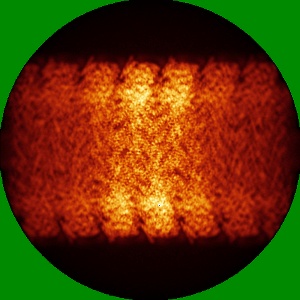
Lipids in biological membranes are thought to be functionally organized, but few experimental tools can probe nanoscale membrane structure. Using brominated lipids as contrast probes for cryo-EM and a model ESCRT-III membrane-remodeling system composed of human CHMP1B and IST1, we observed leaflet-level and protein-localized structural lipid patterns within highly constricted and thinned membrane nanotubes. These nanotubes differed markedly from protein-free, flat bilayers in leaflet thickness, lipid diffusion rates and lipid compositional and conformational asymmetries. Simulations and cryo-EM imaging of brominated stearoyl-docosahexanenoyl-phosphocholine showed how a pair of phenylalanine residues scored the outer leaflet with a helical hydrophobic defect where polyunsaturated docosahexaenoyl tails accumulated at the bilayer surface. Combining cryo-EM of halogenated lipids with molecular dynamics thus enables new characterizations of the composition and structure of membranes on molecular length scales.
Lipids in biological membranes are thought to be functionally organized, but few experimental tools can probe nanoscale membrane structure. Using brominated lipids as contrast probes for cryo-EM and a model ESCRT-III membrane-remodeling system composed of human CHMP1B and IST1, we observed leaflet-level and protein-localized structural lipid patterns within highly constricted and thinned membrane nanotubes. These nanotubes differed markedly from protein-free, flat bilayers in leaflet thickness, lipid diffusion rates and lipid compositional and conformational asymmetries. Simulations and cryo-EM imaging of brominated stearoyl-docosahexanenoyl-phosphocholine showed how a pair of phenylalanine residues scored the outer leaflet with a helical hydrophobic defect where polyunsaturated docosahexaenoyl tails accumulated at the bilayer surface. Combining cryo-EM of halogenated lipids with molecular dynamics thus enables new characterizations of the composition and structure of membranes on molecular length scales.