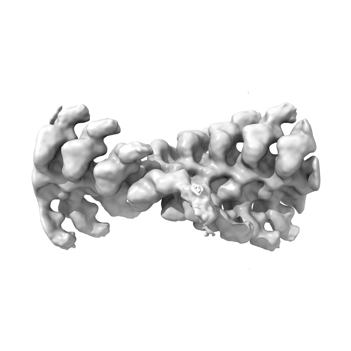
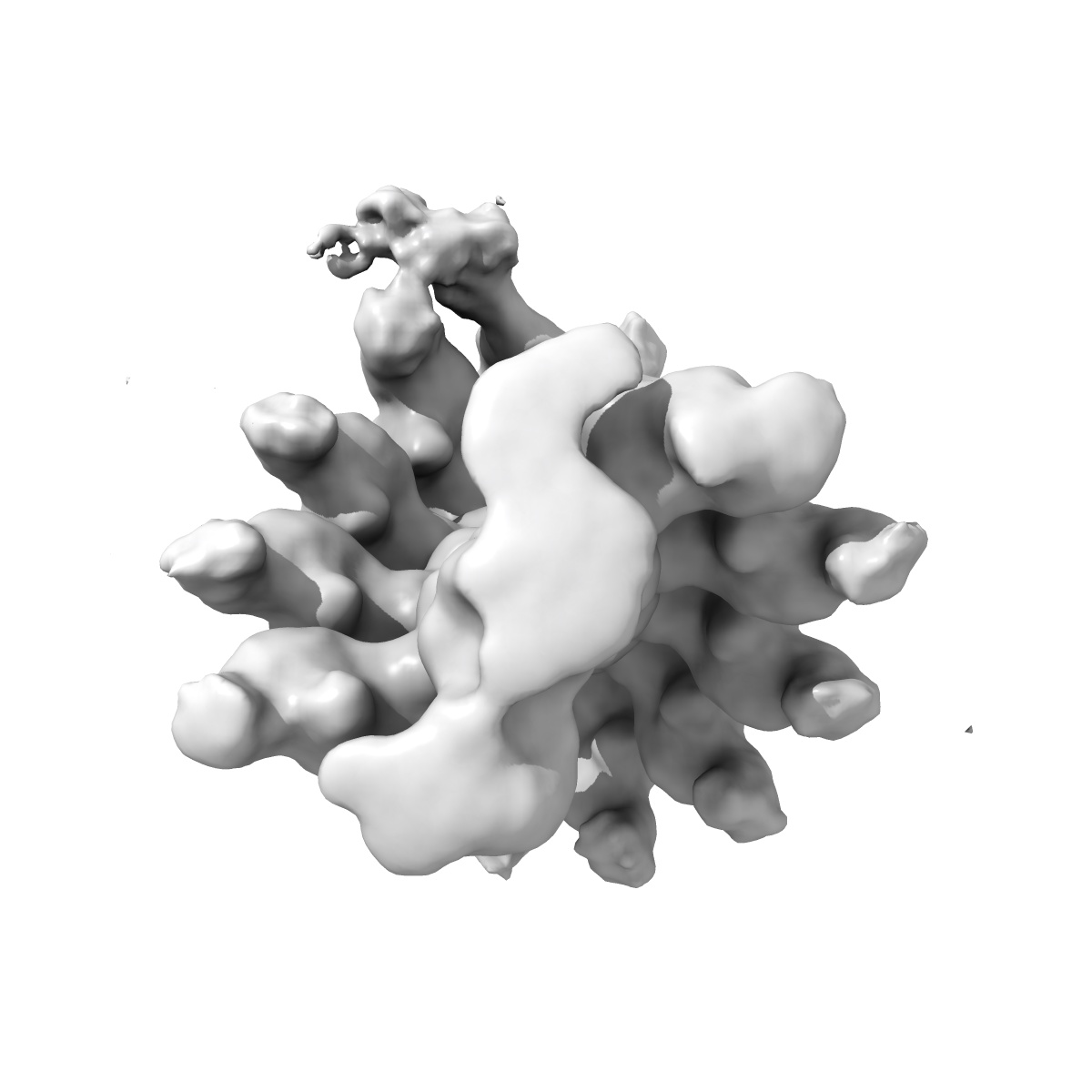
EMD-29646
Acto-HMM complex in ADP-state. Chicken smooth muscle HMM and chicken pectoralis actin
EMD-29646
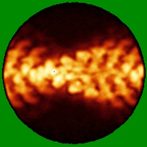
Single-particle19.0 Å
 Deposition: 31/01/2023
Map released: 30/08/2023
Last modified: 30/08/2023
Sample Organism:
Gallus gallus
Sample: Acto-HMM complex - chicken smooth muscle heavy meromyosin in complex with chicken pectoralis muscle actin filament
Fitted models: 8g06
Raw data: EMPIAR-11555
Deposition Authors: Hojjatian A, Liu J, Taylor DW, Daneshparvar N, Trybus KM, Taylor KA
Sample: Acto-HMM complex - chicken smooth muscle heavy meromyosin in complex with chicken pectoralis muscle actin filament
Fitted models: 8g06
Raw data: EMPIAR-11555
Deposition Authors: Hojjatian A, Liu J, Taylor DW, Daneshparvar N, Trybus KM, Taylor KA
Double-headed binding of myosin II to F-actin shows the effect of strain on head structure.
Hojjatian A,
Taylor DW,
Daneshparvar N,
Fagnant PM,
Trybus KM,
Taylor KA
(2023) J Struct Biol , 215 , 107995 - 107995
(2023) J Struct Biol , 215 , 107995 - 107995
Abstract:
Force production in muscle is achieved through the interaction of myosin and actin. Strong binding states in active muscle are associated with Mg·ADP bound to the active site; release of Mg·ADP allows rebinding of ATP and dissociation from actin. Thus, Mg·ADP binding is positioned for adaptation as a force sensor. Mechanical loads on the lever arm can affect the ability of myosin to release Mg·ADP but exactly how this is done is poorly defined. Here we use F-actin decorated with double-headed smooth muscle myosin fragments in the presence of Mg·ADP to visualize the effect of internally supplied tension on the paired lever arms using cryoEM. The interaction of the paired heads with two adjacent actin subunits is predicted to place one lever arm under positive and the other under negative strain. The converter domain is believed to be the most flexible domain within myosin head. Our results, instead, point to the segment of heavy chain between the essential and regulatory light chains as the location of the largest structural change. Moreover, our results suggest no large changes in the myosin coiled coil tail as the locus of strain relief when both heads bind F-actin. The method would be adaptable to double-headed members of the myosin family. We anticipate that the study of actin-myosin interaction using double-headed fragments enables visualization of domains that are typically noisy in decoration with single-headed fragments.
Force production in muscle is achieved through the interaction of myosin and actin. Strong binding states in active muscle are associated with Mg·ADP bound to the active site; release of Mg·ADP allows rebinding of ATP and dissociation from actin. Thus, Mg·ADP binding is positioned for adaptation as a force sensor. Mechanical loads on the lever arm can affect the ability of myosin to release Mg·ADP but exactly how this is done is poorly defined. Here we use F-actin decorated with double-headed smooth muscle myosin fragments in the presence of Mg·ADP to visualize the effect of internally supplied tension on the paired lever arms using cryoEM. The interaction of the paired heads with two adjacent actin subunits is predicted to place one lever arm under positive and the other under negative strain. The converter domain is believed to be the most flexible domain within myosin head. Our results, instead, point to the segment of heavy chain between the essential and regulatory light chains as the location of the largest structural change. Moreover, our results suggest no large changes in the myosin coiled coil tail as the locus of strain relief when both heads bind F-actin. The method would be adaptable to double-headed members of the myosin family. We anticipate that the study of actin-myosin interaction using double-headed fragments enables visualization of domains that are typically noisy in decoration with single-headed fragments.