EMD-30007
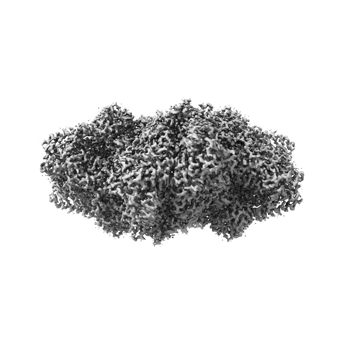
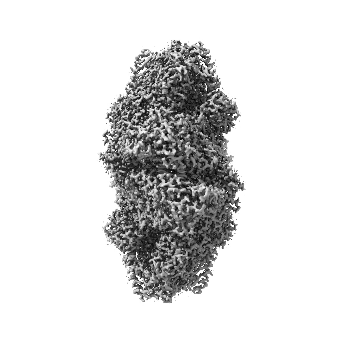
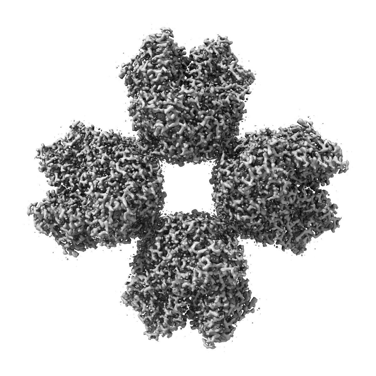
Cryo-EM structure of phosphoketolase from Bifidobacterium longum
EMD-30007
Single-particle2.1 Å
 Deposition: 12/02/2020
Map released: 17/02/2021
Last modified: 29/05/2024
Sample Organism:
Bifidobacterium longum subsp. longum F8
Sample: Phosphoketolase with thiamine-diphophate
Fitted models: 6lxv (Avg. Q-score: 0.763)
Deposition Authors: Nakata K ,
Miyazaki N
,
Miyazaki N
Sample: Phosphoketolase with thiamine-diphophate
Fitted models: 6lxv (Avg. Q-score: 0.763)
Deposition Authors: Nakata K
 ,
Miyazaki N
,
Miyazaki N
High-resolution structure of phosphoketolase from Bifidobacterium longum determined by cryo-EM single-particle analysis.
Nakata K ,
Miyazaki N ,
Yamaguchi H ,
Hirose M ,
Kashiwagi T,
Kutumbarao NHV,
Miyashita O,
Tama F ,
Miyano H,
Mizukoshi T,
Iwasaki K
(2022) J Struct Biol , 214 , 107842 - 107842
,
Miyazaki N ,
Yamaguchi H ,
Hirose M ,
Kashiwagi T,
Kutumbarao NHV,
Miyashita O,
Tama F ,
Miyano H,
Mizukoshi T,
Iwasaki K
(2022) J Struct Biol , 214 , 107842 - 107842
Abstract:
In bifidobacteria, phosphoketolase (PKT) plays a key role in the central hexose fermentation pathway called "bifid shunt." The three-dimensional structure of PKT from Bifidobacterium longum with co-enzyme thiamine diphosphate (ThDpp) was determined at 2.1 Å resolution by cryo-EM single-particle analysis using 196,147 particles to build up the structural model of a PKT octamer related by D4 symmetry. Although the cryo-EM structure of PKT was almost identical to the X-ray crystal structure previously determined at 2.2 Å resolution, several interesting structural features were observed in the cryo-EM structure. Because this structure was solved at relatively high resolution, it was observed that several amino acid residues adopt multiple conformations. Among them, Q546-D547-H548-N549 (the QN-loop) demonstrate the largest structural change, which seems to be related to the enzymatic function of PKT. The QN-loop is at the entrance to the substrate binding pocket. The minor conformer of the QN-loop is similar to the conformation of the QN-loop in the crystal structure. The major conformer is located further from ThDpp than the minor conformer. Interestingly, the major conformer in the cryo-EM structure of PKT resembles the corresponding loop structure of substrate-bound Escherichia coli transketolase. That is, the minor and major conformers may correspond to "closed" and "open" states for substrate access, respectively. Moreover, because of the high-resolution analysis, many water molecules were observed in the cryo-EM structure of PKT. Structural features of the water molecules in the cryo-EM structure are discussed and compared with water molecules observed in the crystal structure.
In bifidobacteria, phosphoketolase (PKT) plays a key role in the central hexose fermentation pathway called "bifid shunt." The three-dimensional structure of PKT from Bifidobacterium longum with co-enzyme thiamine diphosphate (ThDpp) was determined at 2.1 Å resolution by cryo-EM single-particle analysis using 196,147 particles to build up the structural model of a PKT octamer related by D4 symmetry. Although the cryo-EM structure of PKT was almost identical to the X-ray crystal structure previously determined at 2.2 Å resolution, several interesting structural features were observed in the cryo-EM structure. Because this structure was solved at relatively high resolution, it was observed that several amino acid residues adopt multiple conformations. Among them, Q546-D547-H548-N549 (the QN-loop) demonstrate the largest structural change, which seems to be related to the enzymatic function of PKT. The QN-loop is at the entrance to the substrate binding pocket. The minor conformer of the QN-loop is similar to the conformation of the QN-loop in the crystal structure. The major conformer is located further from ThDpp than the minor conformer. Interestingly, the major conformer in the cryo-EM structure of PKT resembles the corresponding loop structure of substrate-bound Escherichia coli transketolase. That is, the minor and major conformers may correspond to "closed" and "open" states for substrate access, respectively. Moreover, because of the high-resolution analysis, many water molecules were observed in the cryo-EM structure of PKT. Structural features of the water molecules in the cryo-EM structure are discussed and compared with water molecules observed in the crystal structure.