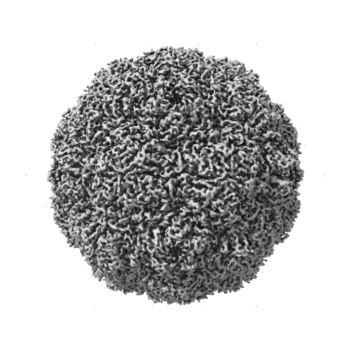
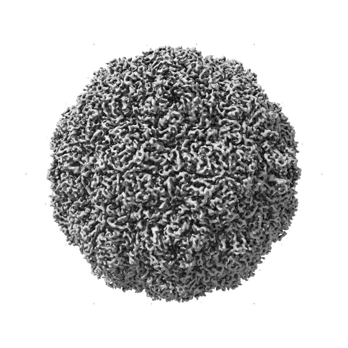
EMD-3137
Multiple capsid-stabilizing protein-RNA and protein-protein interactions revealed in a high-resolution structure of an emerging picornavirus causing neonatal sepsis
EMD-3137
Single-particle4.3 Å
 Deposition: 03/09/2015
Map released: 27/07/2016
Last modified: 10/08/2016
Sample Organism:
Human parechovirus 3
Sample: Human parechovirus 3 capsid structure
Fitted models: 5apm (Avg. Q-score: 0.281)
Raw data: EMPIAR-10033
Deposition Authors: Shakeel S ,
Westerhuis BM,
Domanska A ,
Koning RI ,
Matadeen R,
Koster AJ ,
Bakker AQ,
Beaumont T,
Wolthers KC,
Butcher SJ
,
Westerhuis BM,
Domanska A ,
Koning RI ,
Matadeen R,
Koster AJ ,
Bakker AQ,
Beaumont T,
Wolthers KC,
Butcher SJ
Sample: Human parechovirus 3 capsid structure
Fitted models: 5apm (Avg. Q-score: 0.281)
Raw data: EMPIAR-10033
Deposition Authors: Shakeel S
 ,
Westerhuis BM,
Domanska A ,
Koning RI ,
Matadeen R,
Koster AJ ,
Bakker AQ,
Beaumont T,
Wolthers KC,
Butcher SJ
,
Westerhuis BM,
Domanska A ,
Koning RI ,
Matadeen R,
Koster AJ ,
Bakker AQ,
Beaumont T,
Wolthers KC,
Butcher SJ
Multiple capsid-stabilizing interactions revealed in a high-resolution structure of an emerging picornavirus causing neonatal sepsis
Shakeel S ,
Westerhuis BM,
Domanska A ,
Koning RI ,
Matadeen R,
Koster AJ ,
Beaumont T,
Wolthers KC,
Butcher SJ
(2016) Nat Commun , 7 , 11387
,
Westerhuis BM,
Domanska A ,
Koning RI ,
Matadeen R,
Koster AJ ,
Beaumont T,
Wolthers KC,
Butcher SJ
(2016) Nat Commun , 7 , 11387
Abstract:
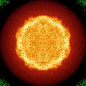
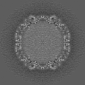
The poorly studied picornavirus, human parechovirus 3 (HPeV3) causes neonatal sepsis with no therapies available. Our 4.3-Å resolution structure of HPeV3 on its own and at 15 Å resolution in complex with human monoclonal antibody Fabs demonstrates the expected picornavirus capsid structure with three distinct features. First, 25% of the HPeV3 RNA genome in 60 sites is highly ordered as confirmed by asymmetric reconstruction, and interacts with conserved regions of the capsid proteins VP1 and VP3. Second, the VP0 N terminus stabilizes the capsid inner surface, in contrast to other picornaviruses where on expulsion as VP4, it forms an RNA translocation channel. Last, VP1's hydrophobic pocket, the binding site for the antipicornaviral drug, pleconaril, is blocked and thus inappropriate for antiviral development. Together, these results suggest a direction for development of neutralizing antibodies, antiviral drugs based on targeting the RNA-protein interactions and dissection of virus assembly on the basis of RNA nucleation.
The poorly studied picornavirus, human parechovirus 3 (HPeV3) causes neonatal sepsis with no therapies available. Our 4.3-Å resolution structure of HPeV3 on its own and at 15 Å resolution in complex with human monoclonal antibody Fabs demonstrates the expected picornavirus capsid structure with three distinct features. First, 25% of the HPeV3 RNA genome in 60 sites is highly ordered as confirmed by asymmetric reconstruction, and interacts with conserved regions of the capsid proteins VP1 and VP3. Second, the VP0 N terminus stabilizes the capsid inner surface, in contrast to other picornaviruses where on expulsion as VP4, it forms an RNA translocation channel. Last, VP1's hydrophobic pocket, the binding site for the antipicornaviral drug, pleconaril, is blocked and thus inappropriate for antiviral development. Together, these results suggest a direction for development of neutralizing antibodies, antiviral drugs based on targeting the RNA-protein interactions and dissection of virus assembly on the basis of RNA nucleation.