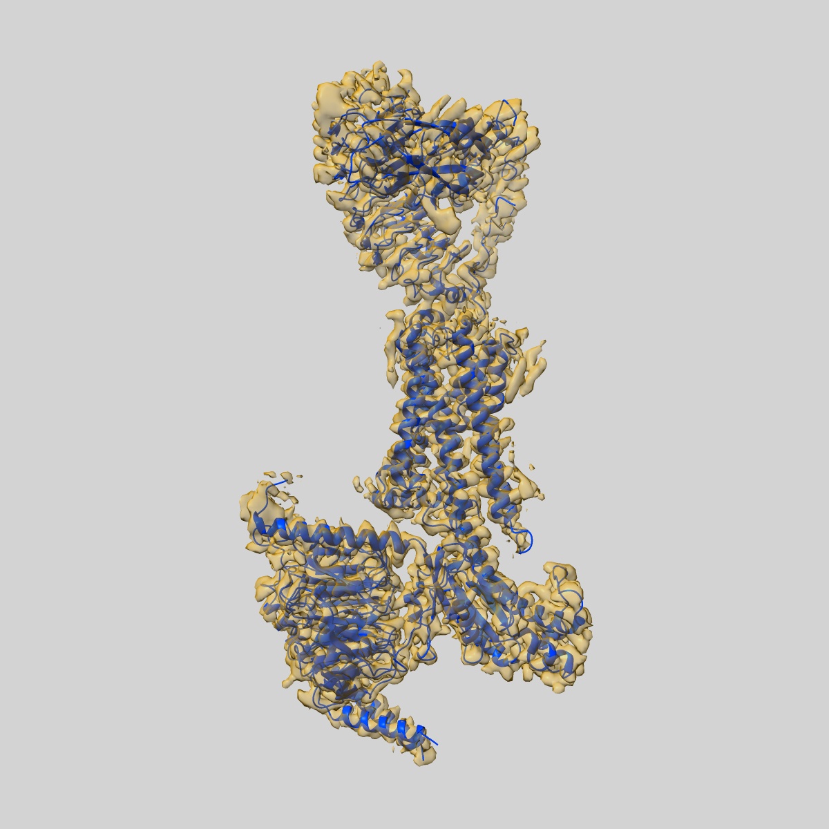
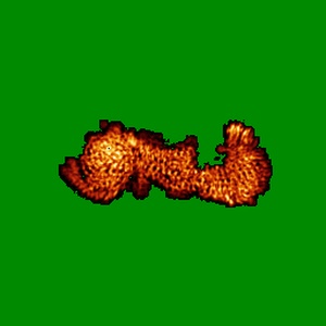
EMD-31597
luteinizing hormone/choriogonadotropin receptor(S277I)-chorionic gonadotropin-Gs-Org43553 complex
EMD-31597
Single-particle3.2 Å
 Deposition: 31/07/2021
Map released: 29/09/2021
Last modified: 09/10/2024
Sample Organism:
Homo sapiens,
Bos taurus,
Rattus norvegicus
Sample: luteinizing hormone/choriogonadotropin receptor(S277I)-chorionic gonadotropin-Gs-Org43553 complex
Fitted models: 7fih (Avg. Q-score: 0.382)
Deposition Authors: Duan J, Xu P
Sample: luteinizing hormone/choriogonadotropin receptor(S277I)-chorionic gonadotropin-Gs-Org43553 complex
Fitted models: 7fih (Avg. Q-score: 0.382)
Deposition Authors: Duan J, Xu P

Structures of full-length glycoprotein hormone receptor signalling complexes.
Duan J,
Xu P ,
Cheng X ,
Mao C ,
Croll T ,
He X ,
Shi J,
Luan X ,
Yin W ,
You E,
Liu Q,
Zhang S ,
Jiang H ,
Zhang Y ,
Jiang Y ,
Xu HE
(2021) Nature , 598 , 688 - 692
,
Cheng X ,
Mao C ,
Croll T ,
He X ,
Shi J,
Luan X ,
Yin W ,
You E,
Liu Q,
Zhang S ,
Jiang H ,
Zhang Y ,
Jiang Y ,
Xu HE
(2021) Nature , 598 , 688 - 692
Abstract:
Luteinizing hormone and chorionic gonadotropin are glycoprotein hormones that are related to follicle-stimulating hormone and thyroid-stimulating hormone1,2. Luteinizing hormone and chorionic gonadotropin are essential to human reproduction and are important therapeutic drugs3-6. They activate the same G-protein-coupled receptor, luteinizing hormone-choriogonadotropin receptor (LHCGR), by binding to the large extracellular domain3. Here we report four cryo-electron microscopy structures of LHCGR: two structures of the wild-type receptor in the inactive and active states; and two structures of the constitutively active mutated receptor. The active structures are bound to chorionic gonadotropin and the stimulatory G protein (Gs), and one of the structures also contains Org43553, an allosteric agonist7. The structures reveal a distinct 'push-and-pull' mechanism of receptor activation, in which the extracellular domain is pushed by the bound hormone and pulled by the extended hinge loop next to the transmembrane domain. A highly conserved 10-residue fragment (P10) from the hinge C-terminal loop at the interface between the extracellular domain and the transmembrane domain functions as a tethered agonist to induce conformational changes in the transmembrane domain and G-protein coupling. Org43553 binds to a pocket of the transmembrane domain and interacts directly with P10, which further stabilizes the active conformation. Together, these structures provide a common model for understanding the signalling of glycoprotein hormone receptors and a basis for drug discovery for endocrine diseases.
Luteinizing hormone and chorionic gonadotropin are glycoprotein hormones that are related to follicle-stimulating hormone and thyroid-stimulating hormone1,2. Luteinizing hormone and chorionic gonadotropin are essential to human reproduction and are important therapeutic drugs3-6. They activate the same G-protein-coupled receptor, luteinizing hormone-choriogonadotropin receptor (LHCGR), by binding to the large extracellular domain3. Here we report four cryo-electron microscopy structures of LHCGR: two structures of the wild-type receptor in the inactive and active states; and two structures of the constitutively active mutated receptor. The active structures are bound to chorionic gonadotropin and the stimulatory G protein (Gs), and one of the structures also contains Org43553, an allosteric agonist7. The structures reveal a distinct 'push-and-pull' mechanism of receptor activation, in which the extracellular domain is pushed by the bound hormone and pulled by the extended hinge loop next to the transmembrane domain. A highly conserved 10-residue fragment (P10) from the hinge C-terminal loop at the interface between the extracellular domain and the transmembrane domain functions as a tethered agonist to induce conformational changes in the transmembrane domain and G-protein coupling. Org43553 binds to a pocket of the transmembrane domain and interacts directly with P10, which further stabilizes the active conformation. Together, these structures provide a common model for understanding the signalling of glycoprotein hormone receptors and a basis for drug discovery for endocrine diseases.