EMD-32264
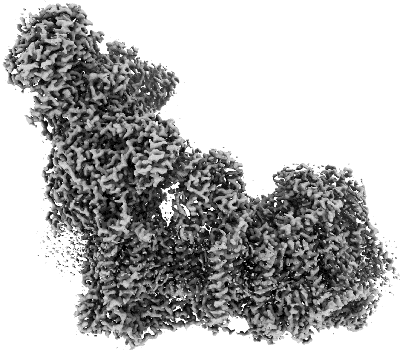
Deactive state CI from Rotenone-NADH dataset, Subclass 2
EMD-32264
Single-particle3.0 Å
 Deposition: 24/11/2021
Map released: 01/02/2023
Last modified: 01/02/2023
Sample Organism:
pig
Sample: Respiratory complex I
Fitted models: 7w2l (Avg. Q-score: 0.57)
Deposition Authors: Gu JK, Yang MJ
Sample: Respiratory complex I
Fitted models: 7w2l (Avg. Q-score: 0.57)
Deposition Authors: Gu JK, Yang MJ
The coupling mechanism of mammalian mitochondrial complex I.

Abstract:
Mammalian respiratory complex I (CI) is a 45-subunit, redox-driven proton pump that generates an electrochemical gradient across the mitochondrial inner membrane to power ATP synthesis in mitochondria. In the present study, we report cryo-electron microscopy structures of CI from Sus scrofa in six treatment conditions at a resolution of 2.4-3.5 Å, in which CI structures of each condition can be classified into two biochemical classes (active or deactive), with a notably higher proportion of active CI particles. These structures illuminate how hydrophobic ubiquinone-10 (Q10) with its long isoprenoid tail is bound and reduced in a narrow Q chamber comprising four different Q10-binding sites. Structural comparisons of active CI structures from our decylubiquinone-NADH and rotenone-NADH datasets reveal that Q10 reduction at site 1 is not coupled to proton pumping in the membrane arm, which might instead be coupled to Q10 oxidation at site 2. Our data overturn the widely accepted previous proposal about the coupling mechanism of CI.
Mammalian respiratory complex I (CI) is a 45-subunit, redox-driven proton pump that generates an electrochemical gradient across the mitochondrial inner membrane to power ATP synthesis in mitochondria. In the present study, we report cryo-electron microscopy structures of CI from Sus scrofa in six treatment conditions at a resolution of 2.4-3.5 Å, in which CI structures of each condition can be classified into two biochemical classes (active or deactive), with a notably higher proportion of active CI particles. These structures illuminate how hydrophobic ubiquinone-10 (Q10) with its long isoprenoid tail is bound and reduced in a narrow Q chamber comprising four different Q10-binding sites. Structural comparisons of active CI structures from our decylubiquinone-NADH and rotenone-NADH datasets reveal that Q10 reduction at site 1 is not coupled to proton pumping in the membrane arm, which might instead be coupled to Q10 oxidation at site 2. Our data overturn the widely accepted previous proposal about the coupling mechanism of CI.