EMD-3227
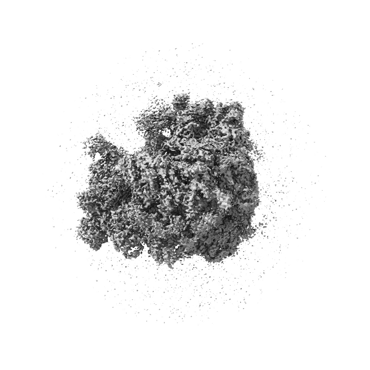
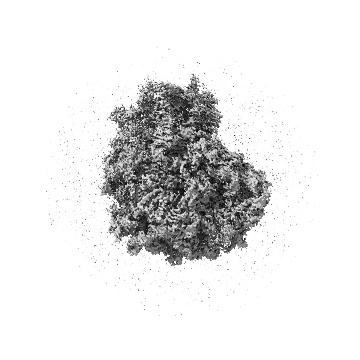
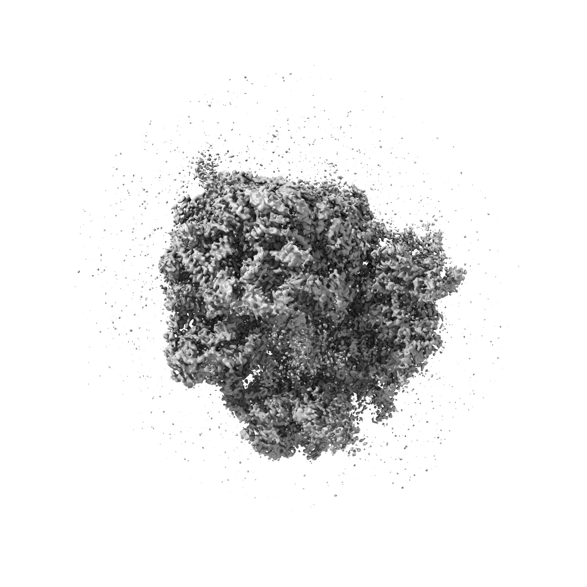
Cryo-EM map of a native 80S-ribosome-eIF-5A complex
EMD-3227
Single-particle3.88 Å
 Deposition: 29/10/2015
Map released: 13/01/2016
Last modified: 16/03/2016
Sample Organism:
Saccharomyces cerevisiae
Sample: Native 80S ribosome-eIF-5A complex from yeast
Fitted models: 5gak (Avg. Q-score: -0.012)
Deposition Authors: Schmidt C ,
Becker T,
Heuer A,
Braunger K,
Shanmuganathan V ,
Pech M,
m Berninghausen O,
Wilson D,
Beckmann R
,
Becker T,
Heuer A,
Braunger K,
Shanmuganathan V ,
Pech M,
m Berninghausen O,
Wilson D,
Beckmann R
Sample: Native 80S ribosome-eIF-5A complex from yeast
Fitted models: 5gak (Avg. Q-score: -0.012)
Deposition Authors: Schmidt C
 ,
Becker T,
Heuer A,
Braunger K,
Shanmuganathan V ,
Pech M,
m Berninghausen O,
Wilson D,
Beckmann R
,
Becker T,
Heuer A,
Braunger K,
Shanmuganathan V ,
Pech M,
m Berninghausen O,
Wilson D,
Beckmann R
Structure of the hypusinylated eukaryotic translation factor eIF-5A bound to the ribosome
Schmidt C ,
Becker T,
Heuer A,
Braunger K,
Shanmuganathan V ,
Pech M,
Berninghausen O,
Wilson DN ,
Beckmann R
(2016) Nucleic Acids Res. , 44 , 1944 - 1951
,
Becker T,
Heuer A,
Braunger K,
Shanmuganathan V ,
Pech M,
Berninghausen O,
Wilson DN ,
Beckmann R
(2016) Nucleic Acids Res. , 44 , 1944 - 1951
Abstract:
During protein synthesis, ribosomes become stalled on polyproline-containing sequences, unless they are rescued in archaea and eukaryotes by the initiation factor 5A (a/eIF-5A) and in bacteria by the homologous protein EF-P. While a structure of EF-P bound to the 70S ribosome exists, structural insight into eIF-5A on the 80S ribosome has been lacking. Here we present a cryo-electron microscopy reconstruction of eIF-5A bound to the yeast 80S ribosome at 3.9 Å resolution. The structure reveals that the unique and functionally essential post-translational hypusine modification reaches toward the peptidyltransferase center of the ribosome, where the hypusine moiety contacts A76 of the CCA-end of the P-site tRNA. These findings would support a model whereby eIF-5A stimulates peptide bond formation on polyproline-stalled ribosomes by stabilizing and orienting the CCA-end of the P-tRNA, rather than by directly contributing to the catalysis.
During protein synthesis, ribosomes become stalled on polyproline-containing sequences, unless they are rescued in archaea and eukaryotes by the initiation factor 5A (a/eIF-5A) and in bacteria by the homologous protein EF-P. While a structure of EF-P bound to the 70S ribosome exists, structural insight into eIF-5A on the 80S ribosome has been lacking. Here we present a cryo-electron microscopy reconstruction of eIF-5A bound to the yeast 80S ribosome at 3.9 Å resolution. The structure reveals that the unique and functionally essential post-translational hypusine modification reaches toward the peptidyltransferase center of the ribosome, where the hypusine moiety contacts A76 of the CCA-end of the P-site tRNA. These findings would support a model whereby eIF-5A stimulates peptide bond formation on polyproline-stalled ribosomes by stabilizing and orienting the CCA-end of the P-tRNA, rather than by directly contributing to the catalysis.