EMD-3256
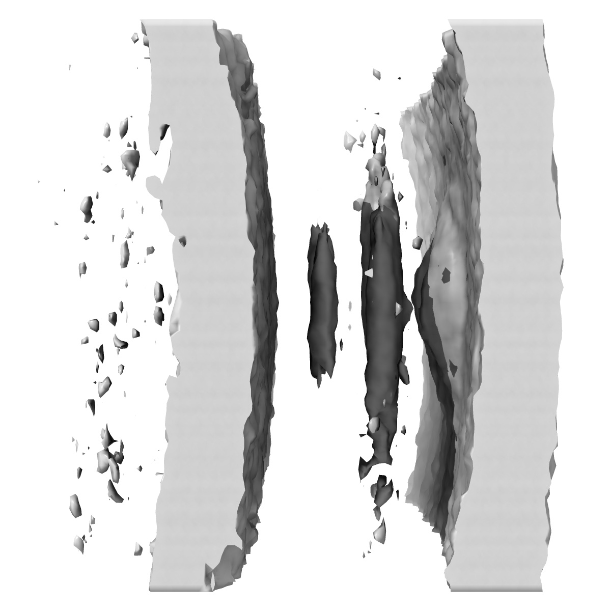
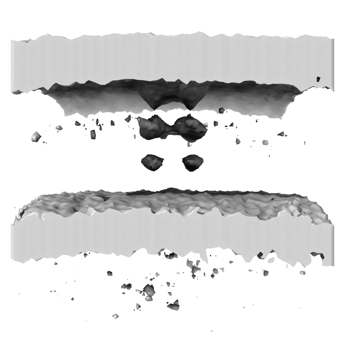
Subtomogram average of a non-piliated type IVa pilus machine in Myxococcus xanthus cells with pilC knockout
EMD-3256
Subtomogram averaging Deposition: 22/11/2015
Map released: 23/03/2016
Last modified: 23/03/2016
Sample Organism:
Myxococcus xanthus
Sample: Myxococcus xanthus DK1622 cells with the pilC gene knockout
Deposition Authors: Chang YW ,
Rettberg LA ,
Treuner-Lange A ,
Iwasa J,
Sogaard-Andersen L ,
Jensen GJ
,
Rettberg LA ,
Treuner-Lange A ,
Iwasa J,
Sogaard-Andersen L ,
Jensen GJ
Sample: Myxococcus xanthus DK1622 cells with the pilC gene knockout
Deposition Authors: Chang YW
 ,
Rettberg LA ,
Treuner-Lange A ,
Iwasa J,
Sogaard-Andersen L ,
Jensen GJ
,
Rettberg LA ,
Treuner-Lange A ,
Iwasa J,
Sogaard-Andersen L ,
Jensen GJ
Architecture of the type IVa pilus machine
Chang YW ,
Rettberg LA ,
Treuner-Lange A ,
Iwasa J,
Sogaard-Andersen L ,
Jensen GJ
(2016) Science , 351
,
Rettberg LA ,
Treuner-Lange A ,
Iwasa J,
Sogaard-Andersen L ,
Jensen GJ
(2016) Science , 351
Abstract:
Type IVa pili are filamentous cell surface structures observed in many bacteria. They pull cells forward by extending, adhering to surfaces, and then retracting. We used cryo-electron tomography of intact Myxococcus xanthus cells to visualize type IVa pili and the protein machine that assembles and retracts them (the type IVa pilus machine, or T4PM) in situ, in both the piliated and nonpiliated states, at a resolution of 3 to 4 nanometers. We found that T4PM comprises an outer membrane pore, four interconnected ring structures in the periplasm and cytoplasm, a cytoplasmic disc and dome, and a periplasmic stem. By systematically imaging mutants lacking defined T4PM proteins or with individual proteins fused to tags, we mapped the locations of all 10 T4PM core components and the minor pilins, thereby providing insights into pilus assembly, structure, and function.
Type IVa pili are filamentous cell surface structures observed in many bacteria. They pull cells forward by extending, adhering to surfaces, and then retracting. We used cryo-electron tomography of intact Myxococcus xanthus cells to visualize type IVa pili and the protein machine that assembles and retracts them (the type IVa pilus machine, or T4PM) in situ, in both the piliated and nonpiliated states, at a resolution of 3 to 4 nanometers. We found that T4PM comprises an outer membrane pore, four interconnected ring structures in the periplasm and cytoplasm, a cytoplasmic disc and dome, and a periplasmic stem. By systematically imaging mutants lacking defined T4PM proteins or with individual proteins fused to tags, we mapped the locations of all 10 T4PM core components and the minor pilins, thereby providing insights into pilus assembly, structure, and function.