EMD-3283
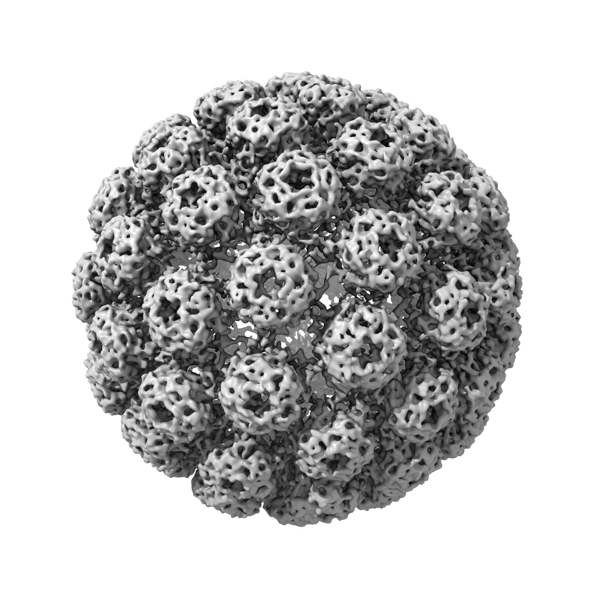
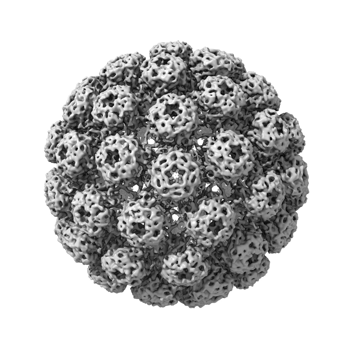
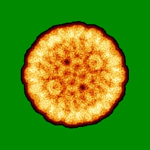
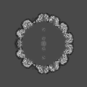
Cryo-EM structure of BK polyomavirus
EMD-3283
Single-particle7.6 Å
 Deposition: 17/12/2015
Map released: 27/04/2016
Last modified: 18/05/2016
Sample Organism:
BK polyomavirus
Sample: BK polyomavirus
Fitted models: 5fua (Avg. Q-score: 0.151)
Deposition Authors: Hurdiss DL ,
Morgan EL ,
Thompson RF,
Prescott EL,
Panou MM,
Macdonald A ,
Ranson NA
,
Morgan EL ,
Thompson RF,
Prescott EL,
Panou MM,
Macdonald A ,
Ranson NA
Sample: BK polyomavirus
Fitted models: 5fua (Avg. Q-score: 0.151)
Deposition Authors: Hurdiss DL
 ,
Morgan EL ,
Thompson RF,
Prescott EL,
Panou MM,
Macdonald A ,
Ranson NA
,
Morgan EL ,
Thompson RF,
Prescott EL,
Panou MM,
Macdonald A ,
Ranson NA
New structural insights into the genome and minor capsid proteins of BK polyomavirus using cryo-electron microscopy
Hurdiss DL ,
Morgan EL ,
Thompson RF,
Prescott EL,
Panou MM,
Macdonald A ,
Ranson NA
(2016) Structure , 24 , 528 - 536
,
Morgan EL ,
Thompson RF,
Prescott EL,
Panou MM,
Macdonald A ,
Ranson NA
(2016) Structure , 24 , 528 - 536
Abstract:
BK polyomavirus is the causative agent of several diseases in transplant patients and the immunosuppressed. In order to better understand the structure and life cycle of BK, we produced infectious virions and VP1-only virus-like particles in cell culture, and determined their three-dimensional structures using cryo-electron microscopy (EM) and single-particle image processing. The resulting 7.6-Å resolution structure of BK and 9.1-Å resolution of the virus-like particles are the highest-resolution cryo-EM structures of any polyomavirus. These structures confirm that the architecture of the major structural protein components of these human polyomaviruses are similar to previous structures from other hosts, but give new insight into the location and role of the enigmatic minor structural proteins, VP2 and VP3. We also observe two shells of electron density, which we attribute to a structurally ordered part of the viral genome, and discrete contacts between this density and both VP1 and the minor capsid proteins.
BK polyomavirus is the causative agent of several diseases in transplant patients and the immunosuppressed. In order to better understand the structure and life cycle of BK, we produced infectious virions and VP1-only virus-like particles in cell culture, and determined their three-dimensional structures using cryo-electron microscopy (EM) and single-particle image processing. The resulting 7.6-Å resolution structure of BK and 9.1-Å resolution of the virus-like particles are the highest-resolution cryo-EM structures of any polyomavirus. These structures confirm that the architecture of the major structural protein components of these human polyomaviruses are similar to previous structures from other hosts, but give new insight into the location and role of the enigmatic minor structural proteins, VP2 and VP3. We also observe two shells of electron density, which we attribute to a structurally ordered part of the viral genome, and discrete contacts between this density and both VP1 and the minor capsid proteins.