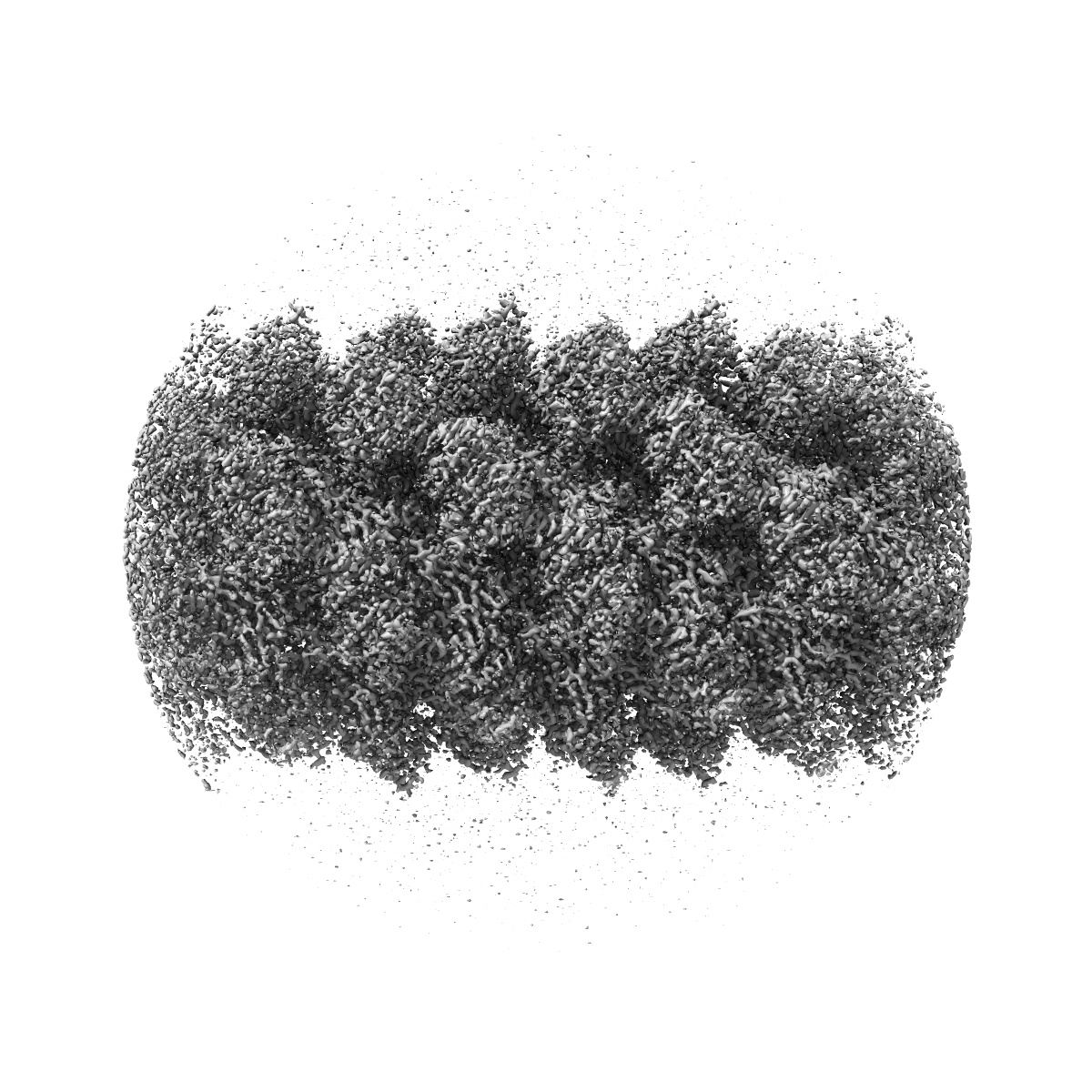
EMD-34681
Cyanophage Pam3 Sheath-tube
EMD-34681
Helical reconstruction3.0 Å
 Deposition: 06/11/2022
Map released: 18/01/2023
Last modified: 03/07/2024
Sample Organism:
uncultured cyanophage
Sample: uncultured cyanophage
Fitted models: 8hdw (Avg. Q-score: 0.516)
Deposition Authors: Yang F ,
Jiang YL,
Zhou CZ
,
Jiang YL,
Zhou CZ
Sample: uncultured cyanophage
Fitted models: 8hdw (Avg. Q-score: 0.516)
Deposition Authors: Yang F
 ,
Jiang YL,
Zhou CZ
,
Jiang YL,
Zhou CZ
Fine structure and assembly pattern of a minimal myophage Pam3.
Yang F ,
Jiang YL,
Zhang JT,
Zhu J ,
Du K,
Yu RC,
Wei ZL,
Kong WW ,
Cui N,
Li WF,
Chen Y ,
Li Q ,
Zhou CZ
(2023) PNAS , 120 , e2213727120 - e2213727120
,
Jiang YL,
Zhang JT,
Zhu J ,
Du K,
Yu RC,
Wei ZL,
Kong WW ,
Cui N,
Li WF,
Chen Y ,
Li Q ,
Zhou CZ
(2023) PNAS , 120 , e2213727120 - e2213727120
Abstract:
The myophage possesses a contractile tail that penetrates its host cell envelope. Except for investigations on the bacteriophage T4 with a rather complicated structure, the assembly pattern and tail contraction mechanism of myophage remain largely unknown. Here, we present the fine structure of a freshwater Myoviridae cyanophage Pam3, which has an icosahedral capsid of ~680 Å in diameter, connected via a three-section neck to an 840-Å-long contractile tail, ending with a three-module baseplate composed of only six protein components. This simplified baseplate consists of a central hub-spike surrounded by six wedge heterotriplexes, to which twelve tail fibers are covalently attached via disulfide bonds in alternating upward and downward configurations. In vitro reduction assays revealed a putative redox-dependent mechanism of baseplate assembly and tail sheath contraction. These findings establish a minimal myophage that might become a user-friendly chassis phage in synthetic biology.
The myophage possesses a contractile tail that penetrates its host cell envelope. Except for investigations on the bacteriophage T4 with a rather complicated structure, the assembly pattern and tail contraction mechanism of myophage remain largely unknown. Here, we present the fine structure of a freshwater Myoviridae cyanophage Pam3, which has an icosahedral capsid of ~680 Å in diameter, connected via a three-section neck to an 840-Å-long contractile tail, ending with a three-module baseplate composed of only six protein components. This simplified baseplate consists of a central hub-spike surrounded by six wedge heterotriplexes, to which twelve tail fibers are covalently attached via disulfide bonds in alternating upward and downward configurations. In vitro reduction assays revealed a putative redox-dependent mechanism of baseplate assembly and tail sheath contraction. These findings establish a minimal myophage that might become a user-friendly chassis phage in synthetic biology.