EMD-35211
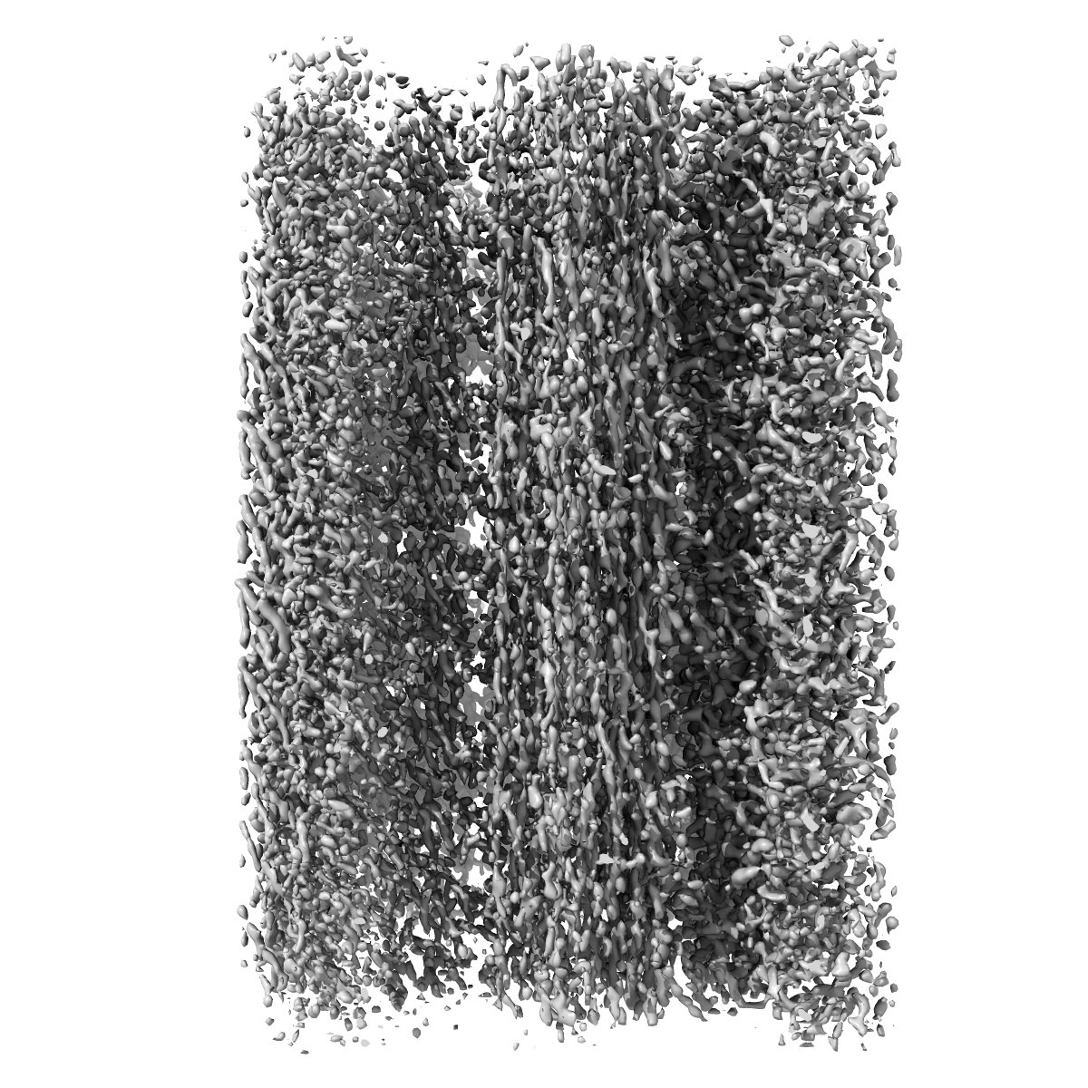
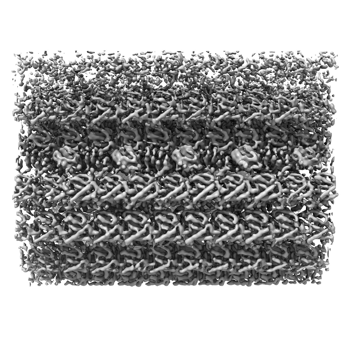
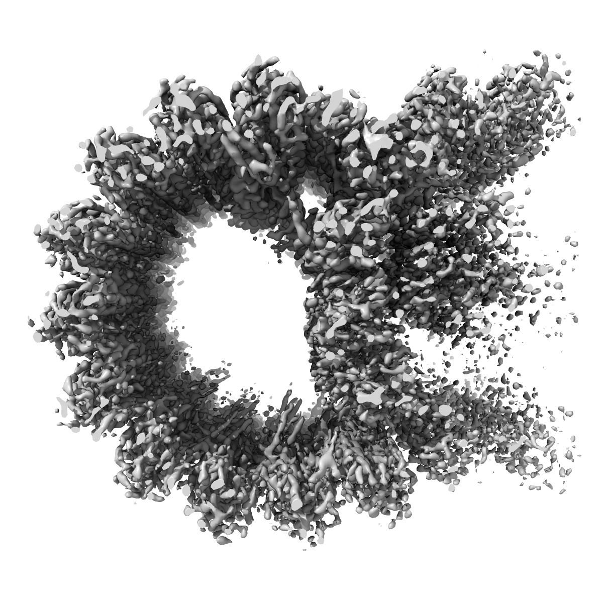
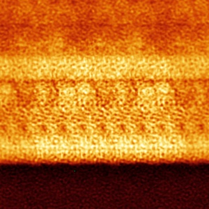
In situ structure of B-tubule of axonemal doublet microtubules in mouse sperm with 16-nm repeat
EMD-35211
Subtomogram averaging6.5 Å
 Deposition: 31/01/2023
Map released: 11/10/2023
Last modified: 06/12/2023
Sample Organism:
Mus musculus
Sample: mouse sperm
Deposition Authors: Zhu Y ,
Tai LH ,
Yin GL ,
Sun F
,
Tai LH ,
Yin GL ,
Sun F
Sample: mouse sperm
Deposition Authors: Zhu Y
 ,
Tai LH ,
Yin GL ,
Sun F
,
Tai LH ,
Yin GL ,
Sun F
In-cell structural insight into the stability of sperm microtubule doublet.
Abstract:
The propulsion for mammalian sperm swimming is generated by flagella beating. Microtubule doublets (DMTs) along with microtubule inner proteins (MIPs) are essential structural blocks of flagella. However, the intricate molecular architecture of intact sperm DMT remains elusive. Here, by in situ cryo-electron tomography, we solved the in-cell structure of mouse sperm DMT at 4.5-7.5 Å resolutions, and built its model with 36 kinds of MIPs in 48 nm periodicity. We identified multiple copies of Tektin5 that reinforce Tektin bundle, and multiple MIPs with different periodicities that anchor the Tektin bundle to tubulin wall. This architecture contributes to a superior stability of A-tubule than B-tubule of DMT, which was revealed by structural comparison of DMTs from the intact and deformed axonemes. Our work provides an overall molecular picture of intact sperm DMT in 48 nm periodicity that is essential to understand the molecular mechanism of sperm motility as well as the related ciliopathies.
The propulsion for mammalian sperm swimming is generated by flagella beating. Microtubule doublets (DMTs) along with microtubule inner proteins (MIPs) are essential structural blocks of flagella. However, the intricate molecular architecture of intact sperm DMT remains elusive. Here, by in situ cryo-electron tomography, we solved the in-cell structure of mouse sperm DMT at 4.5-7.5 Å resolutions, and built its model with 36 kinds of MIPs in 48 nm periodicity. We identified multiple copies of Tektin5 that reinforce Tektin bundle, and multiple MIPs with different periodicities that anchor the Tektin bundle to tubulin wall. This architecture contributes to a superior stability of A-tubule than B-tubule of DMT, which was revealed by structural comparison of DMTs from the intact and deformed axonemes. Our work provides an overall molecular picture of intact sperm DMT in 48 nm periodicity that is essential to understand the molecular mechanism of sperm motility as well as the related ciliopathies.