EMD-35344
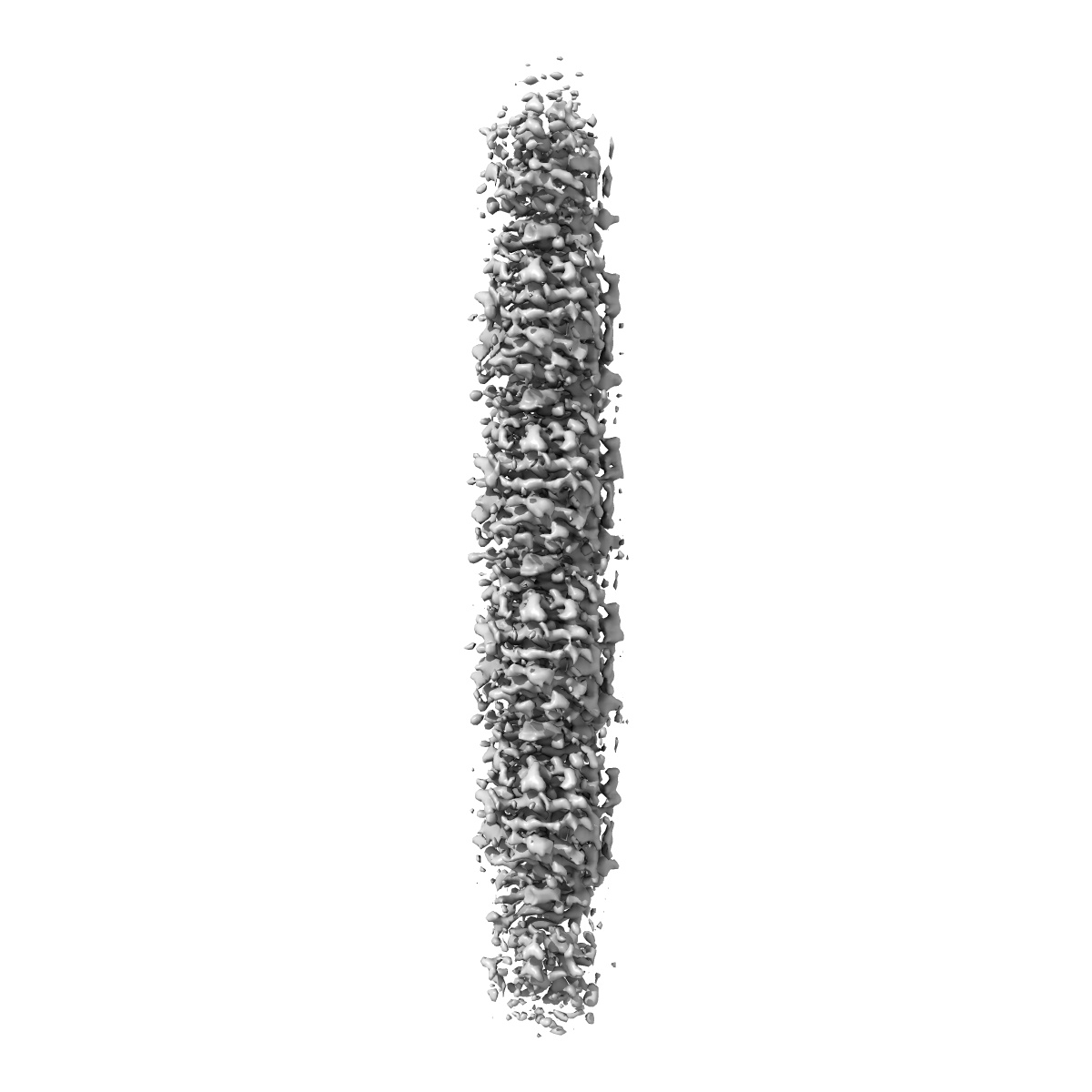
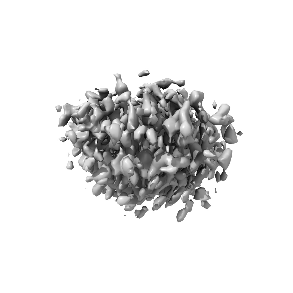
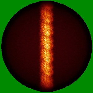
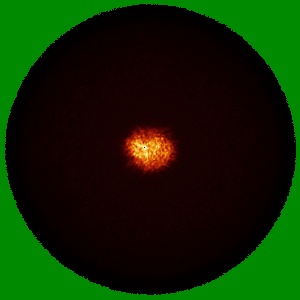
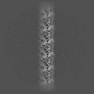
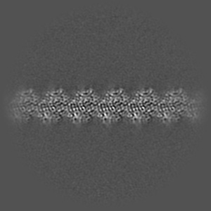
Cryo-EM structure of KpFtsZ single filament
EMD-35344
Helical reconstruction3.03 Å
 Deposition: 10/02/2023
Map released: 02/08/2023
Last modified: 08/05/2024
Sample Organism:
Klebsiella pneumoniae
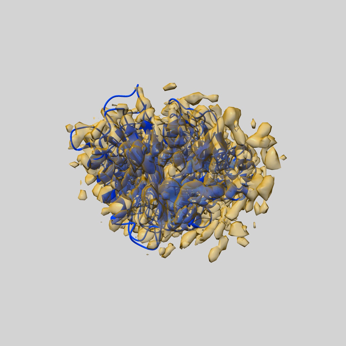
Sample: Cryo-EM structure of KpFtsZ single filament
Fitted models: 8ibn (Avg. Q-score: 0.36)
Raw data: EMPIAR-11426
Deposition Authors: Fujita J ,
Amesaka H ,
Yoshizawa T,
Kuroda N,
Kamimura N,
Hibino K,
Konishi T,
Kato Y,
Hara M,
Inoue T ,
Namba K ,
Tanaka S,
Matsumura H
,
Amesaka H ,
Yoshizawa T,
Kuroda N,
Kamimura N,
Hibino K,
Konishi T,
Kato Y,
Hara M,
Inoue T ,
Namba K ,
Tanaka S,
Matsumura H
Sample: Cryo-EM structure of KpFtsZ single filament
Fitted models: 8ibn (Avg. Q-score: 0.36)
Raw data: EMPIAR-11426
Deposition Authors: Fujita J
 ,
Amesaka H ,
Yoshizawa T,
Kuroda N,
Kamimura N,
Hibino K,
Konishi T,
Kato Y,
Hara M,
Inoue T ,
Namba K ,
Tanaka S,
Matsumura H
,
Amesaka H ,
Yoshizawa T,
Kuroda N,
Kamimura N,
Hibino K,
Konishi T,
Kato Y,
Hara M,
Inoue T ,
Namba K ,
Tanaka S,
Matsumura H
Structures of a FtsZ single protofilament and a double-helical tube in complex with a monobody.
Fujita J ,
Amesaka H ,
Yoshizawa T,
Hibino K,
Kamimura N,
Kuroda N,
Konishi T,
Kato Y,
Hara M,
Inoue T ,
Namba K ,
Tanaka SI ,
Matsumura H
(2023) Nat Commun , 14 , 4073 - 4073
,
Amesaka H ,
Yoshizawa T,
Hibino K,
Kamimura N,
Kuroda N,
Konishi T,
Kato Y,
Hara M,
Inoue T ,
Namba K ,
Tanaka SI ,
Matsumura H
(2023) Nat Commun , 14 , 4073 - 4073
Abstract:
FtsZ polymerizes into protofilaments to form the Z-ring that acts as a scaffold for accessory proteins during cell division. Structures of FtsZ have been previously solved, but detailed mechanistic insights are lacking. Here, we determine the cryoEM structure of a single protofilament of FtsZ from Klebsiella pneumoniae (KpFtsZ) in a polymerization-preferred conformation. We also develop a monobody (Mb) that binds to KpFtsZ and FtsZ from Escherichia coli without affecting their GTPase activity. Crystal structures of the FtsZ-Mb complexes reveal the Mb binding mode, while addition of Mb in vivo inhibits cell division. A cryoEM structure of a double-helical tube of KpFtsZ-Mb at 2.7 Å resolution shows two parallel protofilaments. Our present study highlights the physiological roles of the conformational changes of FtsZ in treadmilling that regulate cell division.
FtsZ polymerizes into protofilaments to form the Z-ring that acts as a scaffold for accessory proteins during cell division. Structures of FtsZ have been previously solved, but detailed mechanistic insights are lacking. Here, we determine the cryoEM structure of a single protofilament of FtsZ from Klebsiella pneumoniae (KpFtsZ) in a polymerization-preferred conformation. We also develop a monobody (Mb) that binds to KpFtsZ and FtsZ from Escherichia coli without affecting their GTPase activity. Crystal structures of the FtsZ-Mb complexes reveal the Mb binding mode, while addition of Mb in vivo inhibits cell division. A cryoEM structure of a double-helical tube of KpFtsZ-Mb at 2.7 Å resolution shows two parallel protofilaments. Our present study highlights the physiological roles of the conformational changes of FtsZ in treadmilling that regulate cell division.