EMD-35976
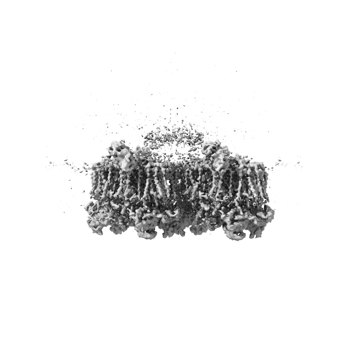
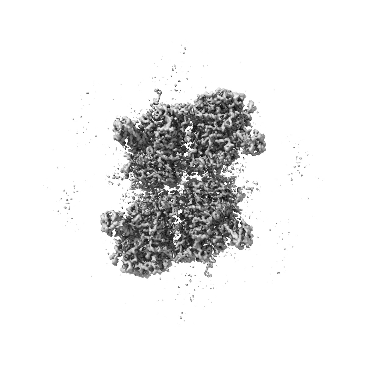
Structure of in situ PSII from P. purpureum lamellae by GisSPA
EMD-35976
Single-particle3.9 Å
 Deposition: 20/04/2023
Map released: 21/06/2023
Last modified: 21/06/2023
Sample Organism:
Porphyridium purpureum
Sample: dimeric PSII complex
Deposition Authors: Zhang X ,
Cheng J
,
Cheng J
Sample: dimeric PSII complex
Deposition Authors: Zhang X
 ,
Cheng J
,
Cheng J
Determining protein structures in cellular lamella at pseudo-atomic resolution by GisSPA.
Abstract:
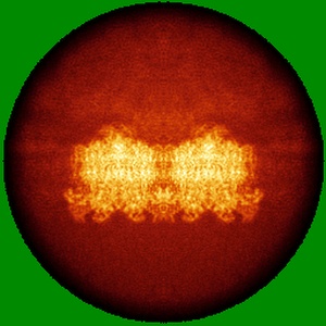
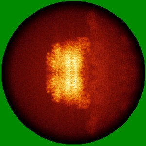
Cryo-electron tomography is a major tool used to study the structure of protein complexes in situ. However, the throughput of tilt-series image data collection is still quite low. Here, we show that GisSPA, a GPU accelerated program, can translationally and rotationally localize the target protein complex in cellular lamellae, as prepared with a focused ion beam, using single cryo-electron microscopy images without tilt-series, and reconstruct the protein complex at near-atomic resolution. GisSPA allows high-throughput data collection without the acquisition of tilt-series images and reconstruction of the tomogram, which is essential for high-resolution reconstruction of asymmetric or low-symmetry protein complexes. We demonstrate the power of GisSPA with 3.4-Å and 3.9-Å resolutions of resolving phycobilisome and tetrameric photosystem II complex structures in cellular lamellae, respectively. In this work, we present GisSPA as a practical tool that facilitates high-resolution in situ protein structure determination.
Cryo-electron tomography is a major tool used to study the structure of protein complexes in situ. However, the throughput of tilt-series image data collection is still quite low. Here, we show that GisSPA, a GPU accelerated program, can translationally and rotationally localize the target protein complex in cellular lamellae, as prepared with a focused ion beam, using single cryo-electron microscopy images without tilt-series, and reconstruct the protein complex at near-atomic resolution. GisSPA allows high-throughput data collection without the acquisition of tilt-series images and reconstruction of the tomogram, which is essential for high-resolution reconstruction of asymmetric or low-symmetry protein complexes. We demonstrate the power of GisSPA with 3.4-Å and 3.9-Å resolutions of resolving phycobilisome and tetrameric photosystem II complex structures in cellular lamellae, respectively. In this work, we present GisSPA as a practical tool that facilitates high-resolution in situ protein structure determination.