EMD-36845
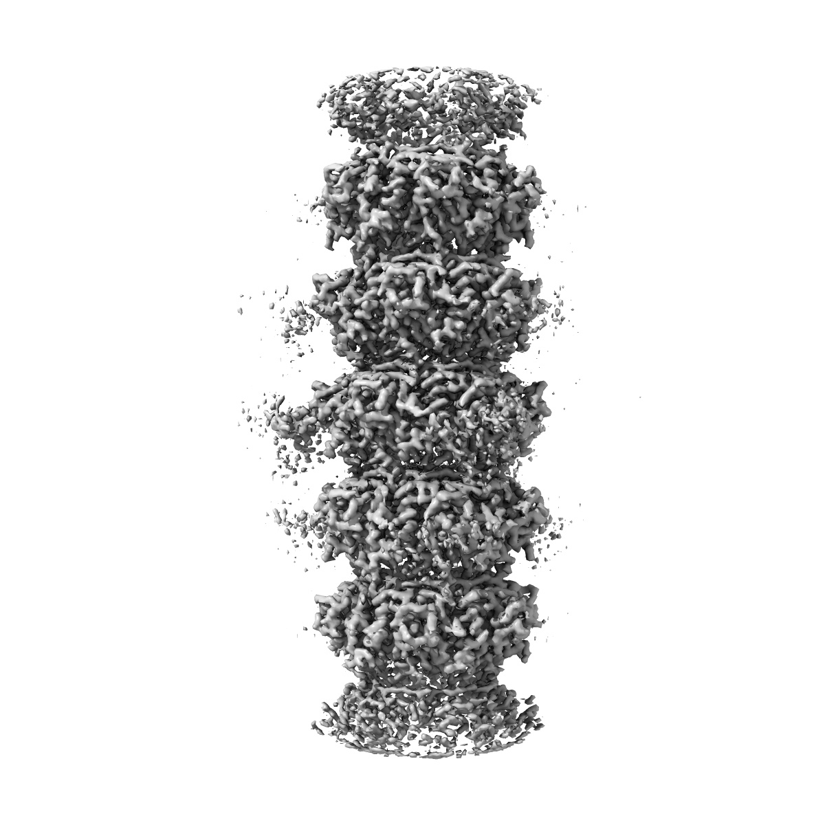
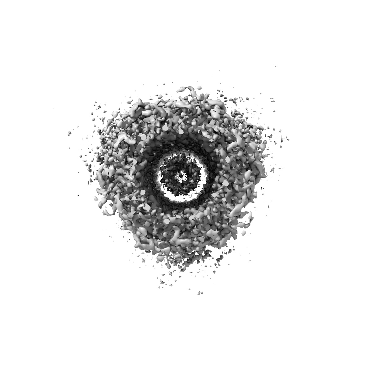
Structure of the bacteriophage lambda tail tube
EMD-36845
Single-particle3.48 Å
 Deposition: 14/07/2023
Map released: 15/11/2023
Last modified: 17/01/2024
Sample Organism:
Escherichia phage Lambda
Sample: Escherichia phage Lambda
Fitted models: 8k36 (Avg. Q-score: 0.54)
Deposition Authors: Xiao H ,
Tan L ,
Cheng LP ,
Liu HR
,
Tan L ,
Cheng LP ,
Liu HR
Sample: Escherichia phage Lambda
Fitted models: 8k36 (Avg. Q-score: 0.54)
Deposition Authors: Xiao H
 ,
Tan L ,
Cheng LP ,
Liu HR
,
Tan L ,
Cheng LP ,
Liu HR
Structure of the siphophage neck-Tail complex suggests that conserved tail tip proteins facilitate receptor binding and tail assembly.
Xiao H ,
Tan L ,
Tan Z,
Zhang Y,
Chen W,
Li X,
Song J,
Cheng L,
Liu H
(2023) PLoS Biol , 21 , e3002441 - e3002441
,
Tan L ,
Tan Z,
Zhang Y,
Chen W,
Li X,
Song J,
Cheng L,
Liu H
(2023) PLoS Biol , 21 , e3002441 - e3002441
Abstract:
Siphophages have a long, flexible, and noncontractile tail that connects to the capsid through a neck. The phage tail is essential for host cell recognition and virus-host cell interactions; moreover, it serves as a channel for genome delivery during infection. However, the in situ high-resolution structure of the neck-tail complex of siphophages remains unknown. Here, we present the structure of the siphophage lambda "wild type," the most widely used, laboratory-adapted fiberless mutant. The neck-tail complex comprises a channel formed by stacked 12-fold and hexameric rings and a 3-fold symmetrical tip. The interactions among DNA and a total of 246 tail protein molecules forming the tail and neck have been characterized. Structural comparisons of the tail tips, the most diversified region across the lambda and other long-tailed phages or tail-like machines, suggest that their tail tip contains conserved domains, which facilitate tail assembly, receptor binding, cell adsorption, and DNA retaining/releasing. These domains are distributed in different tail tip proteins in different phages or tail-like machines. The side tail fibers are not required for the phage particle to orient itself vertically to the surface of the host cell during attachment.
Siphophages have a long, flexible, and noncontractile tail that connects to the capsid through a neck. The phage tail is essential for host cell recognition and virus-host cell interactions; moreover, it serves as a channel for genome delivery during infection. However, the in situ high-resolution structure of the neck-tail complex of siphophages remains unknown. Here, we present the structure of the siphophage lambda "wild type," the most widely used, laboratory-adapted fiberless mutant. The neck-tail complex comprises a channel formed by stacked 12-fold and hexameric rings and a 3-fold symmetrical tip. The interactions among DNA and a total of 246 tail protein molecules forming the tail and neck have been characterized. Structural comparisons of the tail tips, the most diversified region across the lambda and other long-tailed phages or tail-like machines, suggest that their tail tip contains conserved domains, which facilitate tail assembly, receptor binding, cell adsorption, and DNA retaining/releasing. These domains are distributed in different tail tip proteins in different phages or tail-like machines. The side tail fibers are not required for the phage particle to orient itself vertically to the surface of the host cell during attachment.