EMD-39569
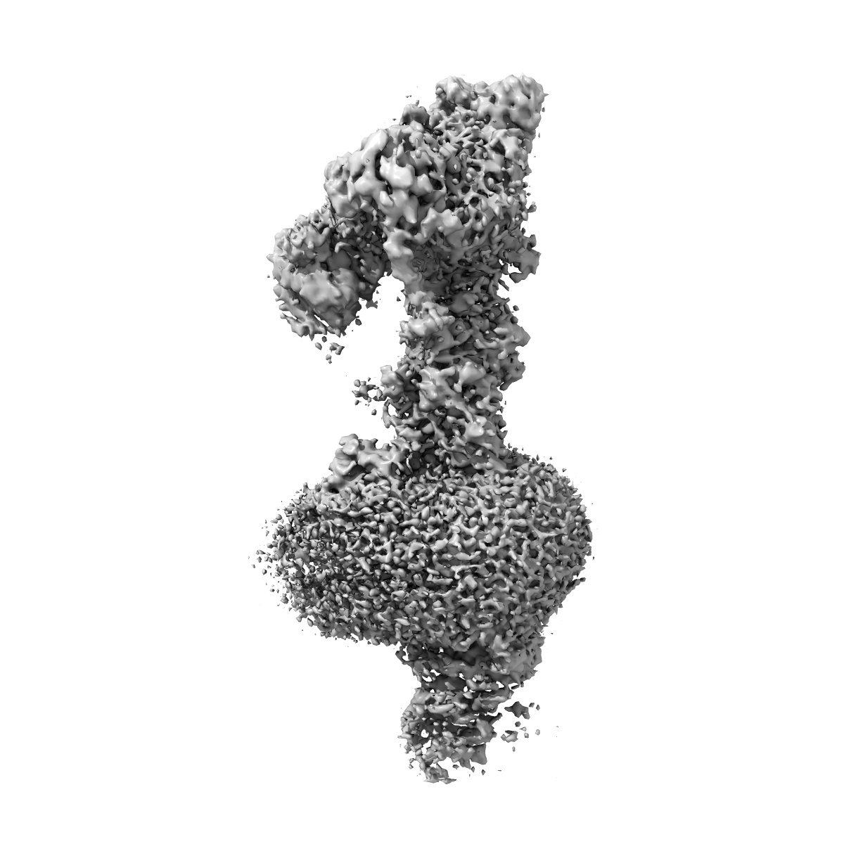
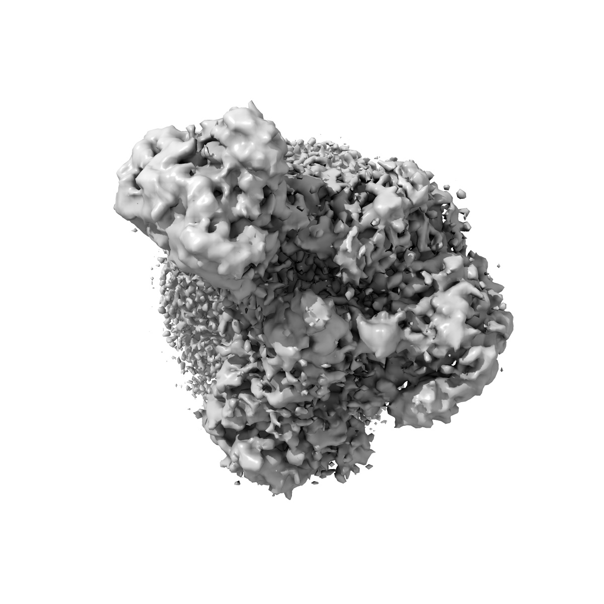
Cryo-EM structure of the dystrophin glycoprotein complex
EMD-39569
Single-particle3.2 Å
 Deposition: 25/03/2024
Map released: 11/12/2024
Last modified: 25/12/2024
Sample Organism:
Mus musculus
Sample: dystrophin glycoprotein complex, DGC
Deposition Authors: Wu JP ,
Yan Z ,
Wan L
,
Yan Z ,
Wan L
Sample: dystrophin glycoprotein complex, DGC
Deposition Authors: Wu JP
 ,
Yan Z ,
Wan L
,
Yan Z ,
Wan L
Structure and assembly of the dystrophin glycoprotein complex.
Abstract:
The dystrophin glycoprotein complex (DGC) has a crucial role in maintaining cell membrane stability and integrity by connecting the intracellular cytoskeleton with the surrounding extracellular matrix1-3. Dysfunction of dystrophin and its associated proteins results in muscular dystrophy, a disorder characterized by progressive muscle weakness and degeneration4,5. Despite the important roles of the DGC in physiology and pathology, its structural details remain largely unknown, hindering a comprehensive understanding of its assembly and function. Here we isolated the native DGC from mouse skeletal muscle and obtained its high-resolution structure. Our findings unveil a markedly divergent structure from the previous model of DGC assembly. Specifically, on the extracellular side, β-, γ- and δ-sarcoglycans co-fold to form a specialized, extracellular tower-like structure, which has a central role in complex assembly by providing binding sites for α-sarcoglycan and dystroglycan. In the transmembrane region, sarcoglycans and sarcospan flank and stabilize the single transmembrane helix of dystroglycan, rather than forming a subcomplex as previously proposed6-8. On the intracellular side, sarcoglycans and dystroglycan engage in assembly with the dystrophin-dystrobrevin subcomplex through extensive interaction with the ZZ domain of dystrophin. Collectively, these findings enhance our understanding of the structural linkage across the cell membrane and provide a foundation for the molecular interpretation of many muscular dystrophy-related mutations.
The dystrophin glycoprotein complex (DGC) has a crucial role in maintaining cell membrane stability and integrity by connecting the intracellular cytoskeleton with the surrounding extracellular matrix1-3. Dysfunction of dystrophin and its associated proteins results in muscular dystrophy, a disorder characterized by progressive muscle weakness and degeneration4,5. Despite the important roles of the DGC in physiology and pathology, its structural details remain largely unknown, hindering a comprehensive understanding of its assembly and function. Here we isolated the native DGC from mouse skeletal muscle and obtained its high-resolution structure. Our findings unveil a markedly divergent structure from the previous model of DGC assembly. Specifically, on the extracellular side, β-, γ- and δ-sarcoglycans co-fold to form a specialized, extracellular tower-like structure, which has a central role in complex assembly by providing binding sites for α-sarcoglycan and dystroglycan. In the transmembrane region, sarcoglycans and sarcospan flank and stabilize the single transmembrane helix of dystroglycan, rather than forming a subcomplex as previously proposed6-8. On the intracellular side, sarcoglycans and dystroglycan engage in assembly with the dystrophin-dystrobrevin subcomplex through extensive interaction with the ZZ domain of dystrophin. Collectively, these findings enhance our understanding of the structural linkage across the cell membrane and provide a foundation for the molecular interpretation of many muscular dystrophy-related mutations.