EMD-3971
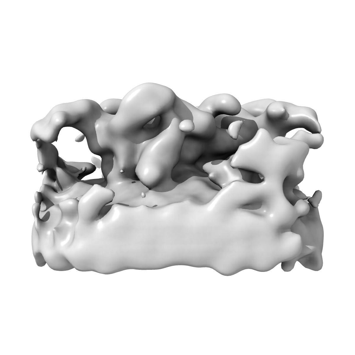
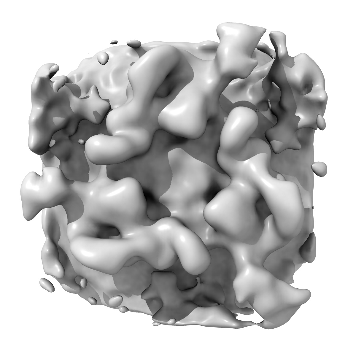
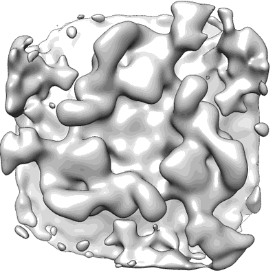
The in situ structure of the Chlamydomonas COPI coat: average from the cis-Golgi region
EMD-3971
Subtomogram averaging24.0 Å
 Deposition: 07/11/2017
Map released: 29/11/2017
Last modified: 29/11/2017
Sample Organism:
Chlamydomonas reinhardtii
Sample: Whole Chlamydomonas cells
Deposition Authors: Bykov YS, Schaffer M, Dodonova SO, Albert S, Plitzko JM, Baumeister W, Engel BD, Briggs JAG
Sample: Whole Chlamydomonas cells
Deposition Authors: Bykov YS, Schaffer M, Dodonova SO, Albert S, Plitzko JM, Baumeister W, Engel BD, Briggs JAG
The structure of the COPI coat determined within the cell.
Bykov YS  ,
Schaffer M ,
Dodonova SO ,
Albert S,
Plitzko JM ,
Baumeister W,
Engel BD ,
Briggs JA
,
Schaffer M ,
Dodonova SO ,
Albert S,
Plitzko JM ,
Baumeister W,
Engel BD ,
Briggs JA
(2017) eLife , 6
 ,
Schaffer M ,
Dodonova SO ,
Albert S,
Plitzko JM ,
Baumeister W,
Engel BD ,
Briggs JA
,
Schaffer M ,
Dodonova SO ,
Albert S,
Plitzko JM ,
Baumeister W,
Engel BD ,
Briggs JA
(2017) eLife , 6
Abstract:
COPI-coated vesicles mediate trafficking within the Golgi apparatus and from the Golgi to the endoplasmic reticulum. The structures of membrane protein coats, including COPI, have been extensively studied with in vitro reconstitution systems using purified components. Previously we have determined a complete structural model of the in vitro reconstituted COPI coat (Dodonova et al., 2017). Here, we applied cryo-focused ion beam milling, cryo-electron tomography and subtomogram averaging to determine the native structure of the COPI coat within vitrified Chlamydomonas reinhardtii cells. The native algal structure resembles the in vitro mammalian structure, but additionally reveals cargo bound beneath β'-COP. We find that all coat components disassemble simultaneously and relatively rapidly after budding. Structural analysis in situ, maintaining Golgi topology, shows that vesicles change their size, membrane thickness, and cargo content as they progress from cis to trans, but the structure of the coat machinery remains constant.
COPI-coated vesicles mediate trafficking within the Golgi apparatus and from the Golgi to the endoplasmic reticulum. The structures of membrane protein coats, including COPI, have been extensively studied with in vitro reconstitution systems using purified components. Previously we have determined a complete structural model of the in vitro reconstituted COPI coat (Dodonova et al., 2017). Here, we applied cryo-focused ion beam milling, cryo-electron tomography and subtomogram averaging to determine the native structure of the COPI coat within vitrified Chlamydomonas reinhardtii cells. The native algal structure resembles the in vitro mammalian structure, but additionally reveals cargo bound beneath β'-COP. We find that all coat components disassemble simultaneously and relatively rapidly after budding. Structural analysis in situ, maintaining Golgi topology, shows that vesicles change their size, membrane thickness, and cargo content as they progress from cis to trans, but the structure of the coat machinery remains constant.