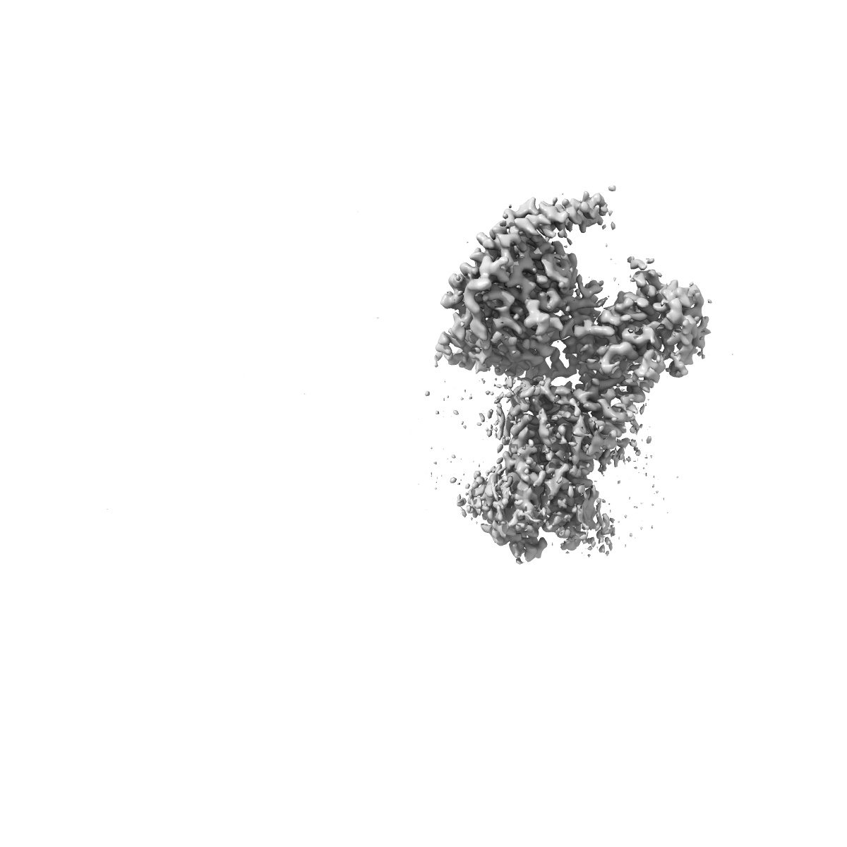
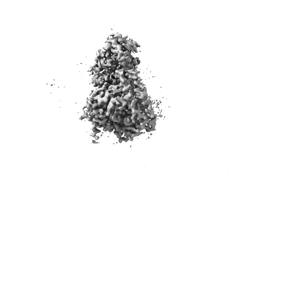
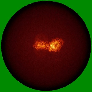
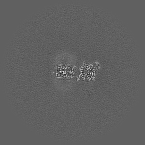
EMD-40109
CryoEM structure of beta-2-adrenergic receptor in complex with Gs heterotrimer, 5 sec after GTP addition (#14 of 20)
EMD-40109
Single-particle3.1 Å
 Deposition: 16/03/2023
Map released: 06/03/2024
Last modified: 05/06/2024
Sample Organism:
Homo sapiens
Sample: Complex of beta-2 adrenergic receptor and Gs heterotrimer, 5 sec post GTP treatment
Raw data: EMPIAR-11856
Deposition Authors: Papasergi-Scott MM ,
Skiniotis G
,
Skiniotis G
Sample: Complex of beta-2 adrenergic receptor and Gs heterotrimer, 5 sec post GTP treatment
Raw data: EMPIAR-11856
Deposition Authors: Papasergi-Scott MM
 ,
Skiniotis G
,
Skiniotis G
Time-resolved cryo-EM of G-protein activation by a GPCR.
Papasergi-Scott MM ,
Perez-Hernandez G ,
Batebi H ,
Gao Y ,
Eskici G ,
Seven AB ,
Panova O ,
Hilger D ,
Casiraghi M,
He F ,
Maul L,
Gmeiner P ,
Kobilka BK ,
Hildebrand PW ,
Skiniotis G
(2024) Nature , 629 , 1182 - 1191
,
Perez-Hernandez G ,
Batebi H ,
Gao Y ,
Eskici G ,
Seven AB ,
Panova O ,
Hilger D ,
Casiraghi M,
He F ,
Maul L,
Gmeiner P ,
Kobilka BK ,
Hildebrand PW ,
Skiniotis G
(2024) Nature , 629 , 1182 - 1191
Abstract:
G-protein-coupled receptors (GPCRs) activate heterotrimeric G proteins by stimulating guanine nucleotide exchange in the Gα subunit1. To visualize this mechanism, we developed a time-resolved cryo-EM approach that examines the progression of ensembles of pre-steady-state intermediates of a GPCR-G-protein complex. By monitoring the transitions of the stimulatory Gs protein in complex with the β2-adrenergic receptor at short sequential time points after GTP addition, we identified the conformational trajectory underlying G-protein activation and functional dissociation from the receptor. Twenty structures generated from sequential overlapping particle subsets along this trajectory, compared to control structures, provide a high-resolution description of the order of main events driving G-protein activation in response to GTP binding. Structural changes propagate from the nucleotide-binding pocket and extend through the GTPase domain, enacting alterations to Gα switch regions and the α5 helix that weaken the G-protein-receptor interface. Molecular dynamics simulations with late structures in the cryo-EM trajectory support that enhanced ordering of GTP on closure of the α-helical domain against the nucleotide-bound Ras-homology domain correlates with α5 helix destabilization and eventual dissociation of the G protein from the GPCR. These findings also highlight the potential of time-resolved cryo-EM as a tool for mechanistic dissection of GPCR signalling events.
G-protein-coupled receptors (GPCRs) activate heterotrimeric G proteins by stimulating guanine nucleotide exchange in the Gα subunit1. To visualize this mechanism, we developed a time-resolved cryo-EM approach that examines the progression of ensembles of pre-steady-state intermediates of a GPCR-G-protein complex. By monitoring the transitions of the stimulatory Gs protein in complex with the β2-adrenergic receptor at short sequential time points after GTP addition, we identified the conformational trajectory underlying G-protein activation and functional dissociation from the receptor. Twenty structures generated from sequential overlapping particle subsets along this trajectory, compared to control structures, provide a high-resolution description of the order of main events driving G-protein activation in response to GTP binding. Structural changes propagate from the nucleotide-binding pocket and extend through the GTPase domain, enacting alterations to Gα switch regions and the α5 helix that weaken the G-protein-receptor interface. Molecular dynamics simulations with late structures in the cryo-EM trajectory support that enhanced ordering of GTP on closure of the α-helical domain against the nucleotide-bound Ras-homology domain correlates with α5 helix destabilization and eventual dissociation of the G protein from the GPCR. These findings also highlight the potential of time-resolved cryo-EM as a tool for mechanistic dissection of GPCR signalling events.