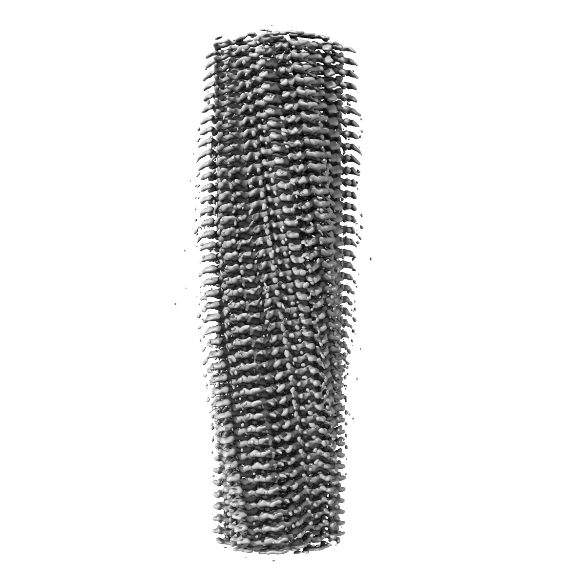
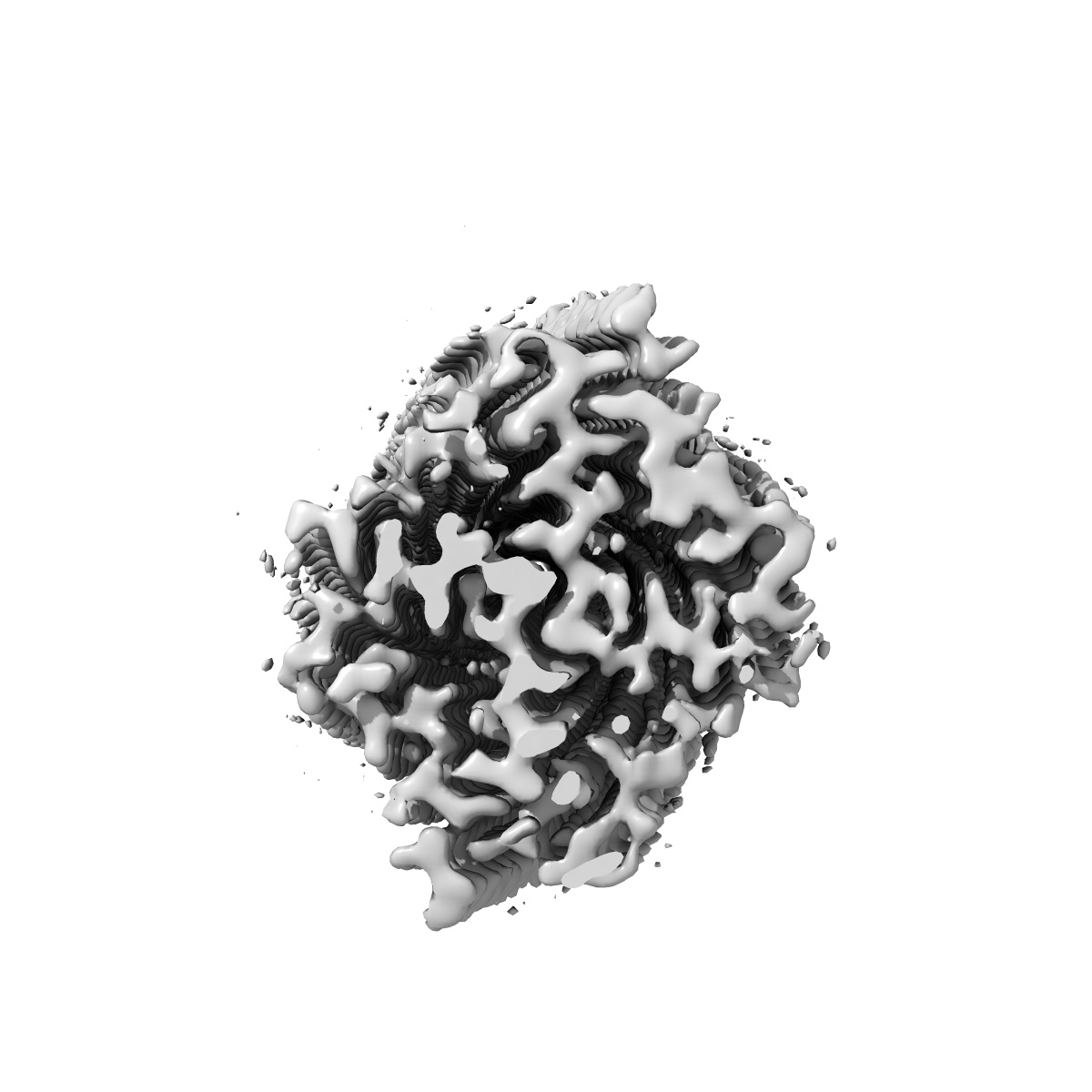
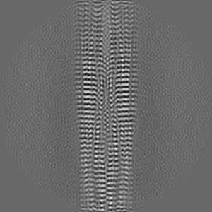
EMD-41198
Tropomyosin-receptor kinase fused gene protein (TRK-fused gene protein; TFG) Low Complexity Domain (residues 237-327) P285L mutant, amyloid fiber
EMD-41198
Helical reconstruction2.59 Å
 Deposition: 06/07/2023
Map released: 20/12/2023
Last modified: 27/12/2023
Sample Organism:
Homo sapiens
Sample: amyloid fibril of protein TFG P285L
Fitted models: 8ter (Avg. Q-score: 0.676)
Deposition Authors: Rosenberg GM ,
Sawaya MR ,
Boyer DR ,
Ge P,
Abskharon R ,
Eisenberg DS
,
Sawaya MR ,
Boyer DR ,
Ge P,
Abskharon R ,
Eisenberg DS
Sample: amyloid fibril of protein TFG P285L
Fitted models: 8ter (Avg. Q-score: 0.676)
Deposition Authors: Rosenberg GM
 ,
Sawaya MR ,
Boyer DR ,
Ge P,
Abskharon R ,
Eisenberg DS
,
Sawaya MR ,
Boyer DR ,
Ge P,
Abskharon R ,
Eisenberg DS
Fibril structures of TFG protein mutants validate the identification of TFG as a disease-related amyloid protein by the IMPAcT method.
Rosenberg GM ,
Abskharon R ,
Boyer DR ,
Ge P,
Sawaya MR ,
Eisenberg DS
(2023) Pnas Nexus , 2 , pgad402 - pgad402
,
Abskharon R ,
Boyer DR ,
Ge P,
Sawaya MR ,
Eisenberg DS
(2023) Pnas Nexus , 2 , pgad402 - pgad402
Abstract:
We previously presented a bioinformatic method for identifying diseases that arise from a mutation in a protein's low-complexity domain that drives the protein into pathogenic amyloid fibrils. One protein so identified was the tropomyosin-receptor kinase-fused gene protein (TRK-fused gene protein or TFG). Mutations in TFG are associated with degenerative neurological conditions. Here, we present experimental evidence that confirms our prediction that these conditions are amyloid-related. We find that the low-complexity domain of TFG containing the disease-related mutations G269V or P285L forms amyloid fibrils, and we determine their structures using cryo-electron microscopy (cryo-EM). These structures are unmistakably amyloid in nature and confirm the propensity of the mutant TFG low-complexity domain to form amyloid fibrils. Also, despite resulting from a pathogenic mutation, the fibril structures bear some similarities to other amyloid structures that are thought to be nonpathogenic and even functional, but there are other factors that support these structures' relevance to disease, including an increased propensity to form amyloid compared with the wild-type sequence, structure-stabilizing influence from the mutant residues themselves, and double-protofilament amyloid cores. Our findings elucidate two potentially disease-relevant structures of a previously unknown amyloid and also show how the structural features of pathogenic amyloid fibrils may not conform to the features commonly associated with pathogenicity.
We previously presented a bioinformatic method for identifying diseases that arise from a mutation in a protein's low-complexity domain that drives the protein into pathogenic amyloid fibrils. One protein so identified was the tropomyosin-receptor kinase-fused gene protein (TRK-fused gene protein or TFG). Mutations in TFG are associated with degenerative neurological conditions. Here, we present experimental evidence that confirms our prediction that these conditions are amyloid-related. We find that the low-complexity domain of TFG containing the disease-related mutations G269V or P285L forms amyloid fibrils, and we determine their structures using cryo-electron microscopy (cryo-EM). These structures are unmistakably amyloid in nature and confirm the propensity of the mutant TFG low-complexity domain to form amyloid fibrils. Also, despite resulting from a pathogenic mutation, the fibril structures bear some similarities to other amyloid structures that are thought to be nonpathogenic and even functional, but there are other factors that support these structures' relevance to disease, including an increased propensity to form amyloid compared with the wild-type sequence, structure-stabilizing influence from the mutant residues themselves, and double-protofilament amyloid cores. Our findings elucidate two potentially disease-relevant structures of a previously unknown amyloid and also show how the structural features of pathogenic amyloid fibrils may not conform to the features commonly associated with pathogenicity.