
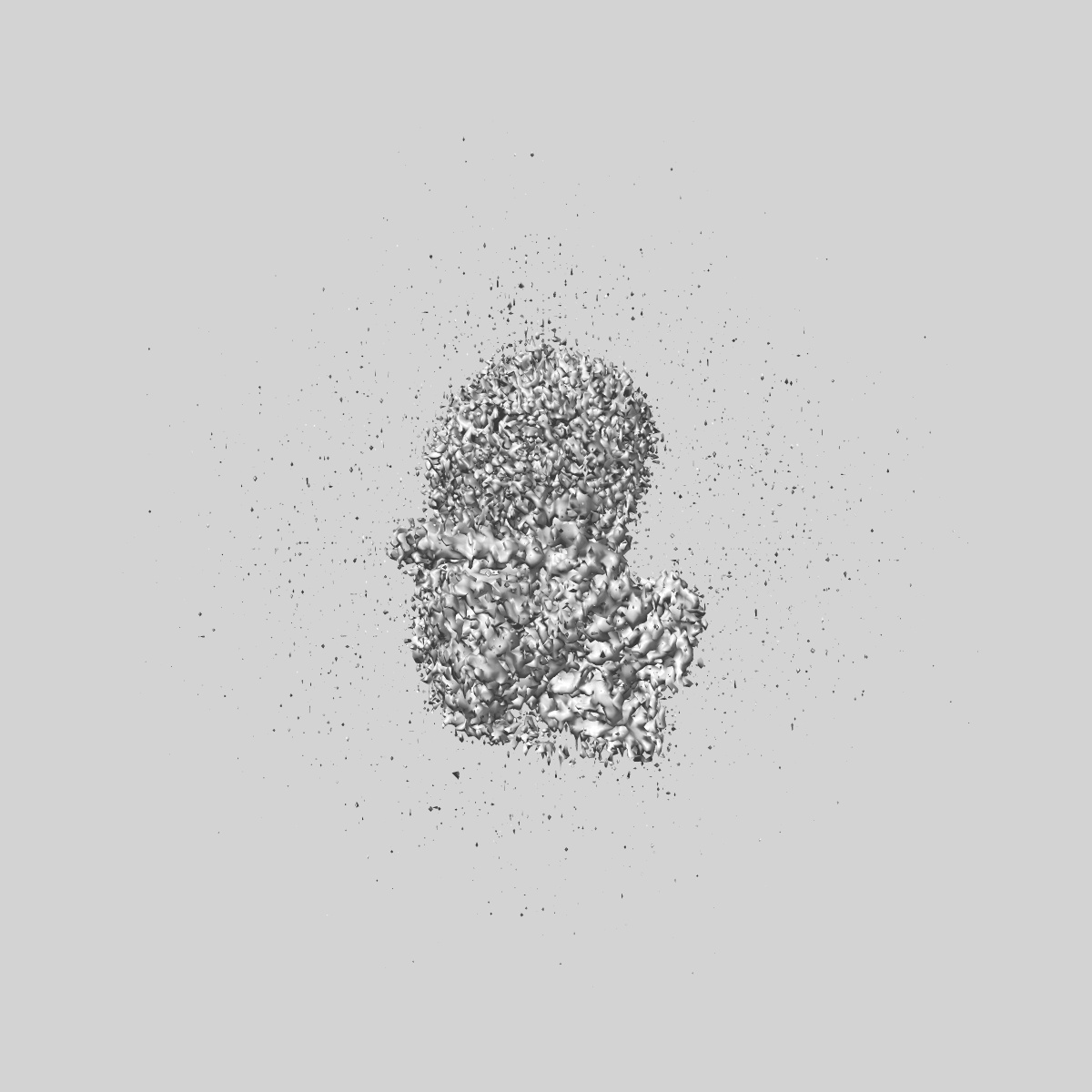
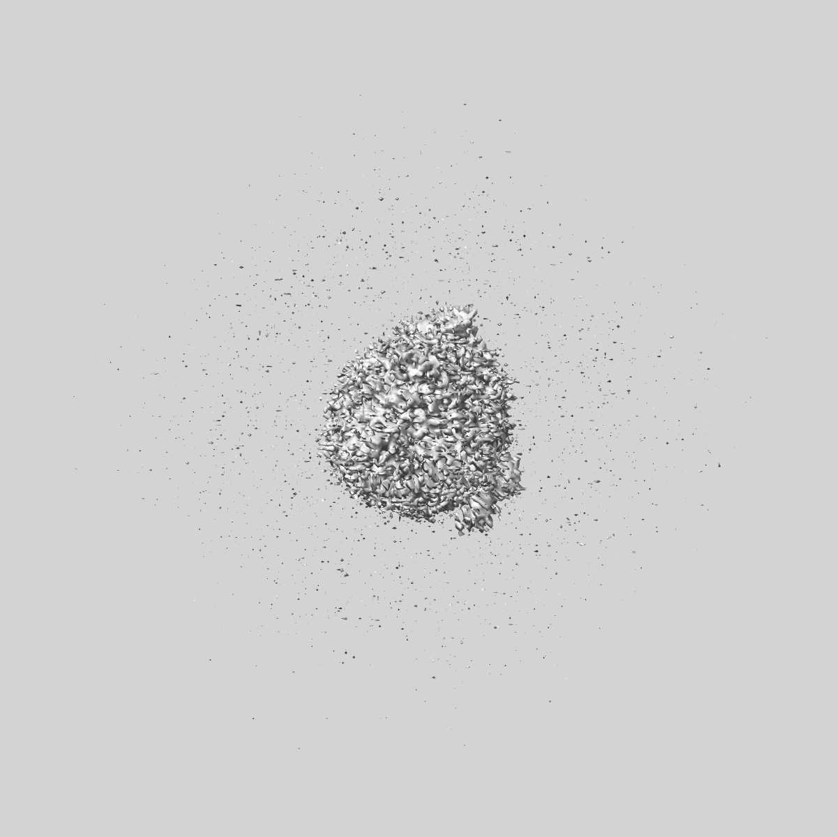
EMD-44550
Human proton sensing receptor GPR68 in complex with miniGs
EMD-44550
Single-particle2.9 Å
 Deposition: 21/04/2024
Map released: 22/01/2025
Last modified: 22/01/2025
Sample Organism:
Homo sapiens,
Lama glama
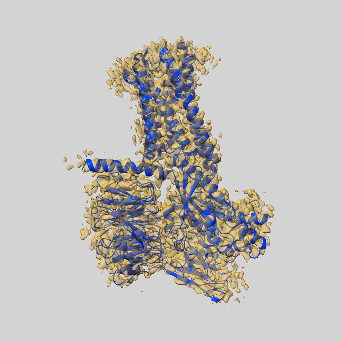
Sample: Complex of GPR68 bound to Gs heterotrimer
Fitted models: 9bhm (Avg. Q-score: 0.456)
Deposition Authors: Howard MK, Hoppe N, Huang XP, Macdonald CB ,
Mehrotra E,
Rockefeller Grimes P,
Zahm AM,
Trinidad DD,
English J,
Coyote-Maestas W,
Manglik A
,
Mehrotra E,
Rockefeller Grimes P,
Zahm AM,
Trinidad DD,
English J,
Coyote-Maestas W,
Manglik A
Sample: Complex of GPR68 bound to Gs heterotrimer
Fitted models: 9bhm (Avg. Q-score: 0.456)
Deposition Authors: Howard MK, Hoppe N, Huang XP, Macdonald CB
 ,
Mehrotra E,
Rockefeller Grimes P,
Zahm AM,
Trinidad DD,
English J,
Coyote-Maestas W,
Manglik A
,
Mehrotra E,
Rockefeller Grimes P,
Zahm AM,
Trinidad DD,
English J,
Coyote-Maestas W,
Manglik A
Molecular basis of proton sensing by G protein-coupled receptors.
Howard MK,
Hoppe N,
Huang XP,
Mitrovic D,
Billesbolle CB,
Macdonald CB ,
Mehrotra E,
Rockefeller Grimes P,
Trinidad DD,
Delemotte L,
English JG,
Coyote-Maestas W,
Manglik A
(2024) Cell
,
Mehrotra E,
Rockefeller Grimes P,
Trinidad DD,
Delemotte L,
English JG,
Coyote-Maestas W,
Manglik A
(2024) Cell
Abstract:
Three proton-sensing G protein-coupled receptors (GPCRs)-GPR4, GPR65, and GPR68-respond to extracellular pH to regulate diverse physiology. How protons activate these receptors is poorly understood. We determined cryogenic-electron microscopy (cryo-EM) structures of each receptor to understand the spatial arrangement of proton-sensing residues. Using deep mutational scanning (DMS), we determined the functional importance of every residue in GPR68 activation by generating ∼9,500 mutants and measuring their effects on signaling and surface expression. Constant-pH molecular dynamics simulations provided insights into the conformational landscape and protonation patterns of key residues. This unbiased approach revealed that, unlike other proton-sensitive channels and receptors, no single site is critical for proton recognition. Instead, a network of titratable residues extends from the extracellular surface to the transmembrane region, converging on canonical motifs to activate proton-sensing GPCRs. Our approach integrating structure, simulations, and unbiased functional interrogation provides a framework for understanding GPCR signaling complexity.
Three proton-sensing G protein-coupled receptors (GPCRs)-GPR4, GPR65, and GPR68-respond to extracellular pH to regulate diverse physiology. How protons activate these receptors is poorly understood. We determined cryogenic-electron microscopy (cryo-EM) structures of each receptor to understand the spatial arrangement of proton-sensing residues. Using deep mutational scanning (DMS), we determined the functional importance of every residue in GPR68 activation by generating ∼9,500 mutants and measuring their effects on signaling and surface expression. Constant-pH molecular dynamics simulations provided insights into the conformational landscape and protonation patterns of key residues. This unbiased approach revealed that, unlike other proton-sensitive channels and receptors, no single site is critical for proton recognition. Instead, a network of titratable residues extends from the extracellular surface to the transmembrane region, converging on canonical motifs to activate proton-sensing GPCRs. Our approach integrating structure, simulations, and unbiased functional interrogation provides a framework for understanding GPCR signaling complexity.