EMD-4461
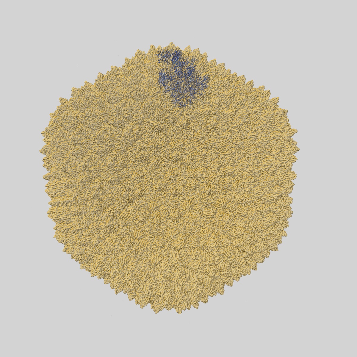
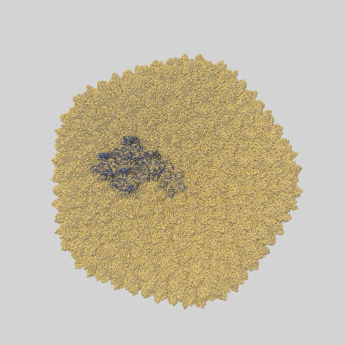
High resolution electron cryo-microscopy structure of the bacteriophage PR772
EMD-4461
Single-particle2.75 Å
 Deposition: 09/12/2018
Map released: 19/06/2019
Last modified: 15/05/2024
Sample Organism:
Enterobacteria phage PR772
Sample: Bacteriophage PR772
Fitted models: 6q5u (Avg. Q-score: 0.574)
Deposition Authors: Narayana Reddy HK, Svenda M
Sample: Bacteriophage PR772
Fitted models: 6q5u (Avg. Q-score: 0.574)
Deposition Authors: Narayana Reddy HK, Svenda M

Electron cryo-microscopy of Bacteriophage PR772 reveals the elusive vertex complex and the capsid architecture.
Abstract:
Bacteriophage PR772, a member of the Tectiviridae family, has a 70 nm diameter icosahedral protein capsid that encapsulates a lipid membrane, dsDNA, and various internal proteins. An icosahedrally averaged CryoEM reconstruction of the wild-type virion and a localized reconstruction of the vertex region reveal the composition and the structure of the vertex complex along with new protein conformations that play a vital role in maintaining the capsid architecture of the virion. The overall resolution of the virion is 2.75 Å, while the resolution of the protein capsid is 2.3 Å. The conventional penta-symmetron formed by the capsomeres is replaced by a large vertex complex in the pseudo T = 25 capsid. All the vertices contain the host-recognition protein, P5; two of these vertices show the presence of the receptor-binding protein, P2. The 3D structure of the vertex complex shows interactions with the viral membrane, indicating a possible mechanism for viral infection.
Bacteriophage PR772, a member of the Tectiviridae family, has a 70 nm diameter icosahedral protein capsid that encapsulates a lipid membrane, dsDNA, and various internal proteins. An icosahedrally averaged CryoEM reconstruction of the wild-type virion and a localized reconstruction of the vertex region reveal the composition and the structure of the vertex complex along with new protein conformations that play a vital role in maintaining the capsid architecture of the virion. The overall resolution of the virion is 2.75 Å, while the resolution of the protein capsid is 2.3 Å. The conventional penta-symmetron formed by the capsomeres is replaced by a large vertex complex in the pseudo T = 25 capsid. All the vertices contain the host-recognition protein, P5; two of these vertices show the presence of the receptor-binding protein, P2. The 3D structure of the vertex complex shows interactions with the viral membrane, indicating a possible mechanism for viral infection.