EMD-4539
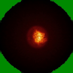
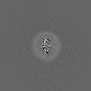
Nanodisc reconstituted human ABCB1 in complex with UIC2 fab and taxol
EMD-4539
Single-particle3.6 Å
 Deposition: 08/01/2019
Map released: 27/02/2019
Last modified: 13/11/2024
Sample Organism:
Mus musculus,
Homo sapiens
Sample: Nanodisc reconstituted human ABCB1 in complex with UIC2 Fab and taxol
Fitted models: 6qex (Avg. Q-score: 0.392)
Deposition Authors: Alam A ,
Locher KP
,
Locher KP
Sample: Nanodisc reconstituted human ABCB1 in complex with UIC2 Fab and taxol
Fitted models: 6qex (Avg. Q-score: 0.392)
Deposition Authors: Alam A
 ,
Locher KP
,
Locher KP
Structural insight into substrate and inhibitor discrimination by human P-glycoprotein.
Abstract:
ABCB1, also known as P-glycoprotein, actively extrudes xenobiotic compounds across the plasma membrane of diverse cells, which contributes to cellular drug resistance and interferes with therapeutic drug delivery. We determined the 3.5-angstrom cryo-electron microscopy structure of substrate-bound human ABCB1 reconstituted in lipidic nanodiscs, revealing a single molecule of the chemotherapeutic compound paclitaxel (Taxol) bound in a central, occluded pocket. A second structure of inhibited, human-mouse chimeric ABCB1 revealed two molecules of zosuquidar occupying the same drug-binding pocket. Minor structural differences between substrate- and inhibitor-bound ABCB1 sites are amplified toward the nucleotide-binding domains (NBDs), revealing how the plasticity of the drug-binding site controls the dynamics of the adenosine triphosphate-hydrolyzing NBDs. Ordered cholesterol and phospholipid molecules suggest how the membrane modulates the conformational changes associated with drug binding and transport.
ABCB1, also known as P-glycoprotein, actively extrudes xenobiotic compounds across the plasma membrane of diverse cells, which contributes to cellular drug resistance and interferes with therapeutic drug delivery. We determined the 3.5-angstrom cryo-electron microscopy structure of substrate-bound human ABCB1 reconstituted in lipidic nanodiscs, revealing a single molecule of the chemotherapeutic compound paclitaxel (Taxol) bound in a central, occluded pocket. A second structure of inhibited, human-mouse chimeric ABCB1 revealed two molecules of zosuquidar occupying the same drug-binding pocket. Minor structural differences between substrate- and inhibitor-bound ABCB1 sites are amplified toward the nucleotide-binding domains (NBDs), revealing how the plasticity of the drug-binding site controls the dynamics of the adenosine triphosphate-hydrolyzing NBDs. Ordered cholesterol and phospholipid molecules suggest how the membrane modulates the conformational changes associated with drug binding and transport.