EMD-45602
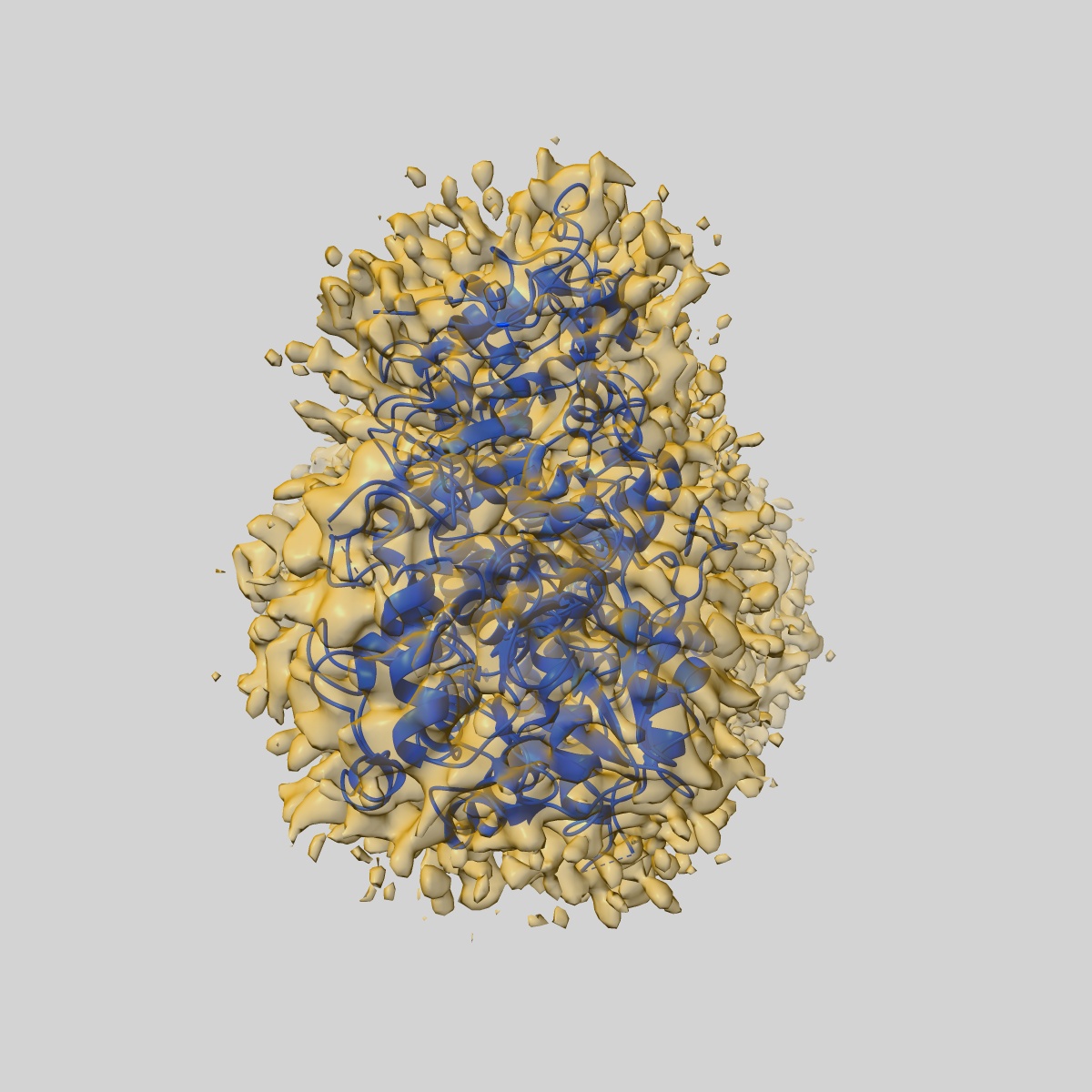
Cryo-EM structure of calcineurin fused beta2 adrenergic receptor in norepinephrine bound inactive state
EMD-45602
Single-particle3.49 Å
 Deposition: 02/07/2024
Map released: 13/11/2024
Last modified: 27/11/2024
Sample Organism:
Homo sapiens,
Mus musculus
Sample: calcineurin fused beta2AR in complex with FKBP12
Fitted models: 9chu (Avg. Q-score: 0.478)
Deposition Authors: Xu J, Chen G, Du Y ,
Kobilka BK
,
Kobilka BK
Sample: calcineurin fused beta2AR in complex with FKBP12
Fitted models: 9chu (Avg. Q-score: 0.478)
Deposition Authors: Xu J, Chen G, Du Y
 ,
Kobilka BK
,
Kobilka BK
Calcineurin-fusion facilitates cryo-EM structure determination of a Family A GPCR.
Xu J,
Chen G,
Wang H,
Cao S,
Heng J,
Deupi X ,
Du Y ,
Kobilka BK
(2024) PNAS , 121 , e2414544121 - e2414544121
,
Du Y ,
Kobilka BK
(2024) PNAS , 121 , e2414544121 - e2414544121
Abstract:
Advances in singe-particle cryo-electron microscopy (cryo-EM) have made it possible to solve the structures of numerous Family A and Family B G protein-coupled receptors (GPCRs) in complex with G proteins and arrestins, as well as several Family C GPCRs. Determination of these structures has been facilitated by the presence of large extramembrane components (such as G protein, arrestin, or Venus flytrap domains) in these complexes that aid in particle alignment during the processing of the cryo-EM data. In contrast, determination of the inactive state structure of Family A GPCRs is more challenging due to the relatively small size of the seven transmembrane domain (7TM) and to the surrounding detergent micelle that, in the absence of other features, make particle alignment impossible. Here, we describe an alternative protein engineering strategy where the heterodimeric protein calcineurin is fused to a GPCR by three points of attachment, the cytoplasmic ends of TM5, TM6, and TM7. This three-point attachment provides a more rigid link with the GPCR transmembrane domain that facilitates particle alignment during data processing, allowing us to determine the structures of the β2 adrenergic receptor (β2AR) in the apo, antagonist-bound, and agonist-bound states. We expect that this fusion strategy may have broad application in cryo-EM structural determination of other Family A GPCRs.
Advances in singe-particle cryo-electron microscopy (cryo-EM) have made it possible to solve the structures of numerous Family A and Family B G protein-coupled receptors (GPCRs) in complex with G proteins and arrestins, as well as several Family C GPCRs. Determination of these structures has been facilitated by the presence of large extramembrane components (such as G protein, arrestin, or Venus flytrap domains) in these complexes that aid in particle alignment during the processing of the cryo-EM data. In contrast, determination of the inactive state structure of Family A GPCRs is more challenging due to the relatively small size of the seven transmembrane domain (7TM) and to the surrounding detergent micelle that, in the absence of other features, make particle alignment impossible. Here, we describe an alternative protein engineering strategy where the heterodimeric protein calcineurin is fused to a GPCR by three points of attachment, the cytoplasmic ends of TM5, TM6, and TM7. This three-point attachment provides a more rigid link with the GPCR transmembrane domain that facilitates particle alignment during data processing, allowing us to determine the structures of the β2 adrenergic receptor (β2AR) in the apo, antagonist-bound, and agonist-bound states. We expect that this fusion strategy may have broad application in cryo-EM structural determination of other Family A GPCRs.