EMD-45672
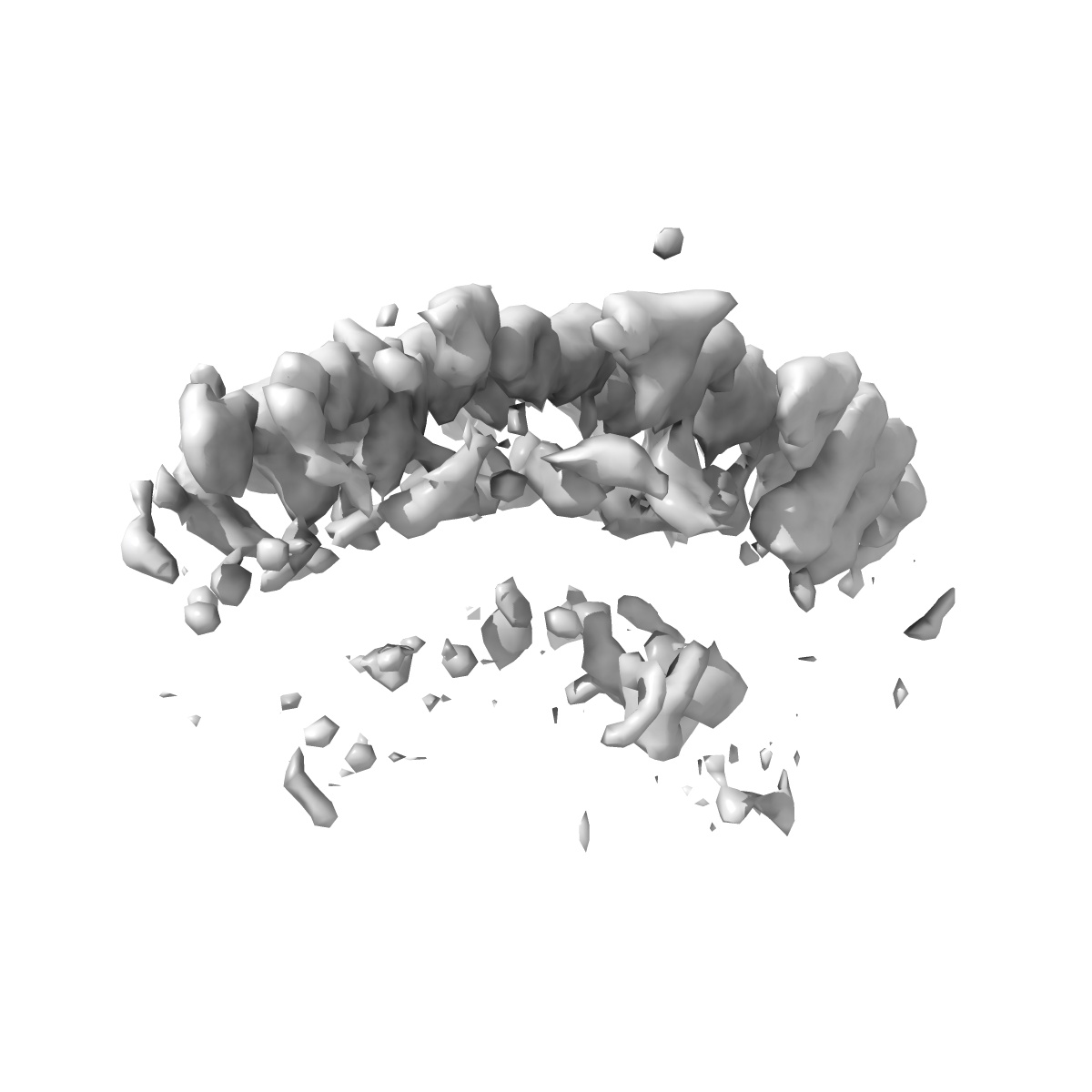
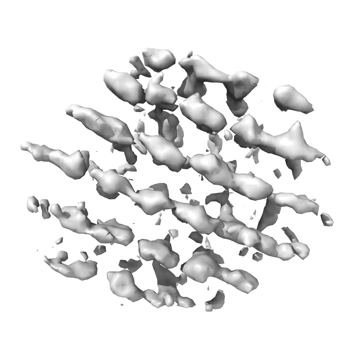
Subtomogram average of the Polar Tube Inner Filament layer from Encephalitozoon intestinalis microsporidian spores
EMD-45672
Subtomogram averaging20.0 Å
 Deposition: 10/07/2024
Map released: 26/02/2025
Last modified: 05/03/2025
Sample Organism:
Encephalitozoon intestinalis
Sample: Encephalitozoon intestinalis microsporidian spores
Deposition Authors: Usmani M ,
Coudray N ,
Bobe D ,
Kopylov M ,
Ekiert DC ,
Bhabha G
,
Coudray N ,
Bobe D ,
Kopylov M ,
Ekiert DC ,
Bhabha G
Sample: Encephalitozoon intestinalis microsporidian spores
Deposition Authors: Usmani M
 ,
Coudray N ,
Bobe D ,
Kopylov M ,
Ekiert DC ,
Bhabha G
,
Coudray N ,
Bobe D ,
Kopylov M ,
Ekiert DC ,
Bhabha G
Cryo-ET reveals the in situ architecture of the polar tube invasion apparatus from microsporidian parasites.
Usmani M ,
Coudray N ,
Riggi M,
Raghu R,
Ramchandani H,
Bobe D ,
Kopylov M ,
Zhong ED,
Iwasa JH,
Ekiert DC ,
Bhabha G
(2024) bioRxiv
,
Coudray N ,
Riggi M,
Raghu R,
Ramchandani H,
Bobe D ,
Kopylov M ,
Zhong ED,
Iwasa JH,
Ekiert DC ,
Bhabha G
(2024) bioRxiv
Abstract:
Microsporidia are divergent fungal pathogens that employ a harpoon-like apparatus called the polar tube (PT) to invade host cells. The PT architecture and its association with neighboring organelles remain poorly understood. Here, we use cryo-electron tomography to investigate the structural cell biology of the PT in dormant spores from the human-infecting microsporidian species, Encephalitozoon intestinalis . Segmentation and subtomogram averaging of the PT reveal at least four layers: two protein-based layers surrounded by a membrane, and filled with a dense core. Regularly spaced protein filaments form the structural skeleton of the PT. Combining cryo-electron tomography with cellular modeling, we propose a model for the 3-dimensional organization of the polaroplast, an organelle that is continuous with the membrane layer that envelops the PT. Our results reveal the ultrastructure of the microsporidian invasion apparatus in situ , laying the foundation for understanding infection mechanisms.
Microsporidia are divergent fungal pathogens that employ a harpoon-like apparatus called the polar tube (PT) to invade host cells. The PT architecture and its association with neighboring organelles remain poorly understood. Here, we use cryo-electron tomography to investigate the structural cell biology of the PT in dormant spores from the human-infecting microsporidian species, Encephalitozoon intestinalis . Segmentation and subtomogram averaging of the PT reveal at least four layers: two protein-based layers surrounded by a membrane, and filled with a dense core. Regularly spaced protein filaments form the structural skeleton of the PT. Combining cryo-electron tomography with cellular modeling, we propose a model for the 3-dimensional organization of the polaroplast, an organelle that is continuous with the membrane layer that envelops the PT. Our results reveal the ultrastructure of the microsporidian invasion apparatus in situ , laying the foundation for understanding infection mechanisms.