EMD-45761
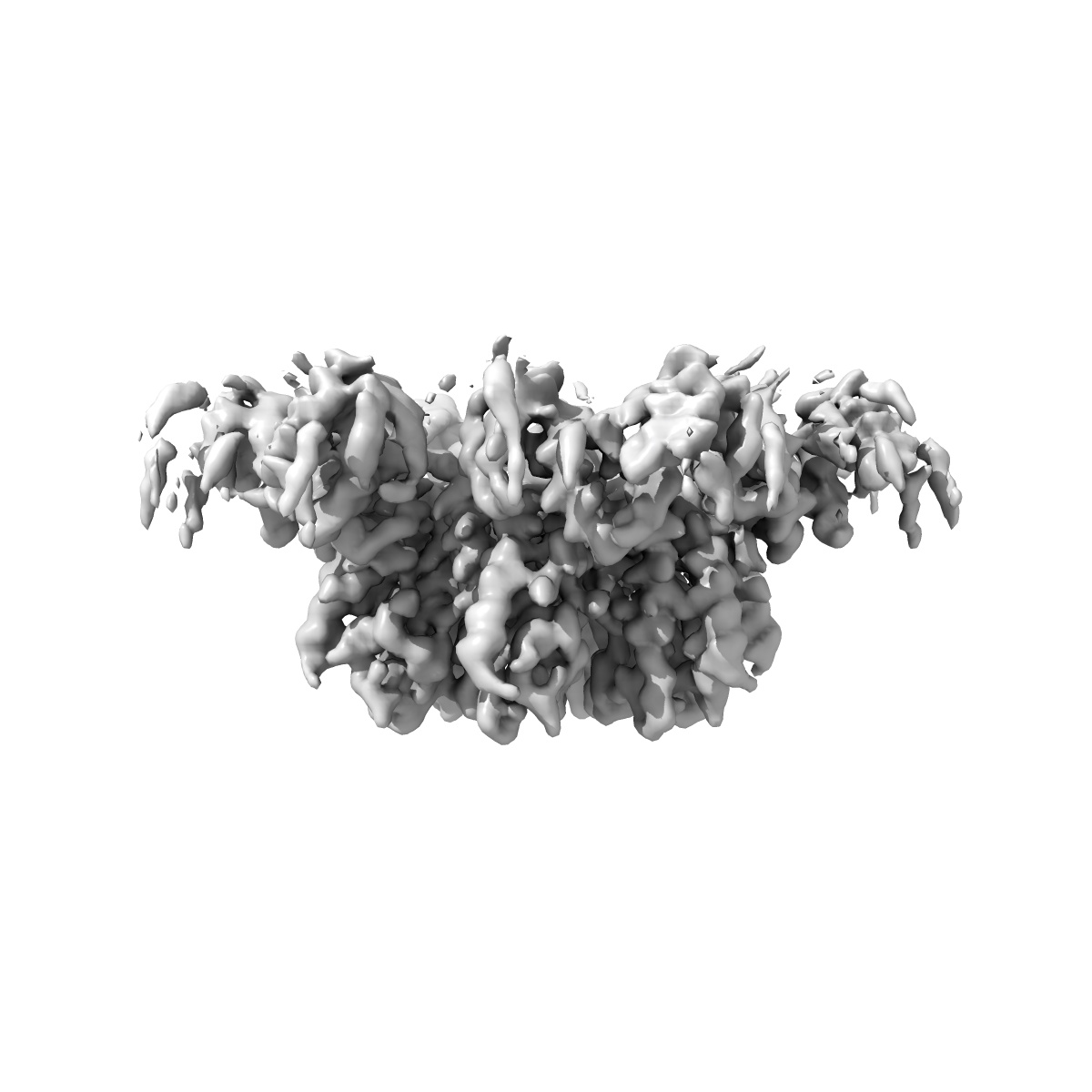
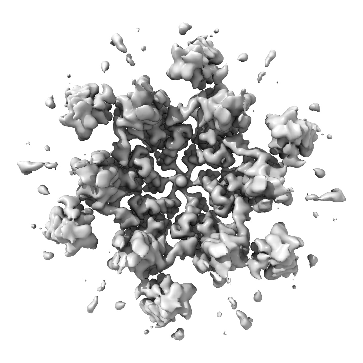
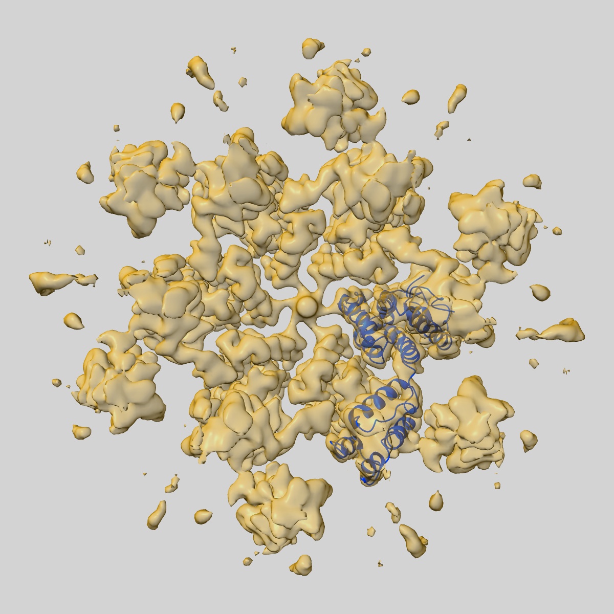
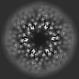
HIV-2 CA hexamer bound with CPSF6 peptide; assembled via liposome templating
EMD-45761
Single-particle3.16 Å
 Deposition: 15/07/2024
Map released: 05/03/2025
Last modified: 05/03/2025
Sample Organism:
Human immunodeficiency virus 2,
Homo sapiens
Sample: HIV-2 capsid protein assembled into a lattice via liposome templating and then bound with CPSF6 peptide.
Fitted models: 9cnv
Deposition Authors: Freniere C ,
Cook M ,
Xiong Y
,
Cook M ,
Xiong Y
Sample: HIV-2 capsid protein assembled into a lattice via liposome templating and then bound with CPSF6 peptide.
Fitted models: 9cnv
Deposition Authors: Freniere C
 ,
Cook M ,
Xiong Y
,
Cook M ,
Xiong Y
Structural insights into HIV-2 CA lattice formation and FG-pocket binding revealed by single-particle cryo-EM.
Abstract:
One of the striking features of human immunodeficiency virus (HIV) is the capsid, a fullerene cone comprised of pleomorphic capsid protein (CA) that shields the viral genome and recruits cofactors. Despite significant advances in understanding the mechanisms of HIV-1 CA assembly and host factor interactions, HIV-2 CA assembly remains poorly understood. By templating the assembly of HIV-2 CA on functionalized liposomes, we report high-resolution structures of the HIV-2 CA lattice, including both CA hexamers and pentamers, alone and with peptides of host phenylalanine-glycine (FG)-motif proteins Nup153 and CPSF6. While the overall fold and mode of FG-peptide binding is conserved with HIV-1, this study reveals distinctive features of the HIV-2 CA lattice, including differing structural character at regions of host factor interactions and divergence in the mechanism of formation of CA hexamers and pentamers. This study extends our understanding of HIV capsids and highlights an approach facilitating the study of lentiviral capsid biology.
One of the striking features of human immunodeficiency virus (HIV) is the capsid, a fullerene cone comprised of pleomorphic capsid protein (CA) that shields the viral genome and recruits cofactors. Despite significant advances in understanding the mechanisms of HIV-1 CA assembly and host factor interactions, HIV-2 CA assembly remains poorly understood. By templating the assembly of HIV-2 CA on functionalized liposomes, we report high-resolution structures of the HIV-2 CA lattice, including both CA hexamers and pentamers, alone and with peptides of host phenylalanine-glycine (FG)-motif proteins Nup153 and CPSF6. While the overall fold and mode of FG-peptide binding is conserved with HIV-1, this study reveals distinctive features of the HIV-2 CA lattice, including differing structural character at regions of host factor interactions and divergence in the mechanism of formation of CA hexamers and pentamers. This study extends our understanding of HIV capsids and highlights an approach facilitating the study of lentiviral capsid biology.