EMD-45866
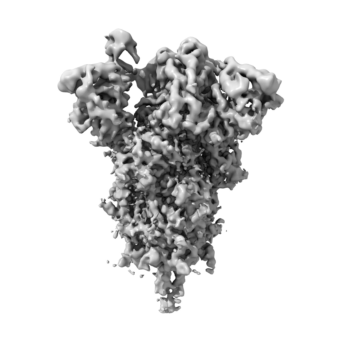
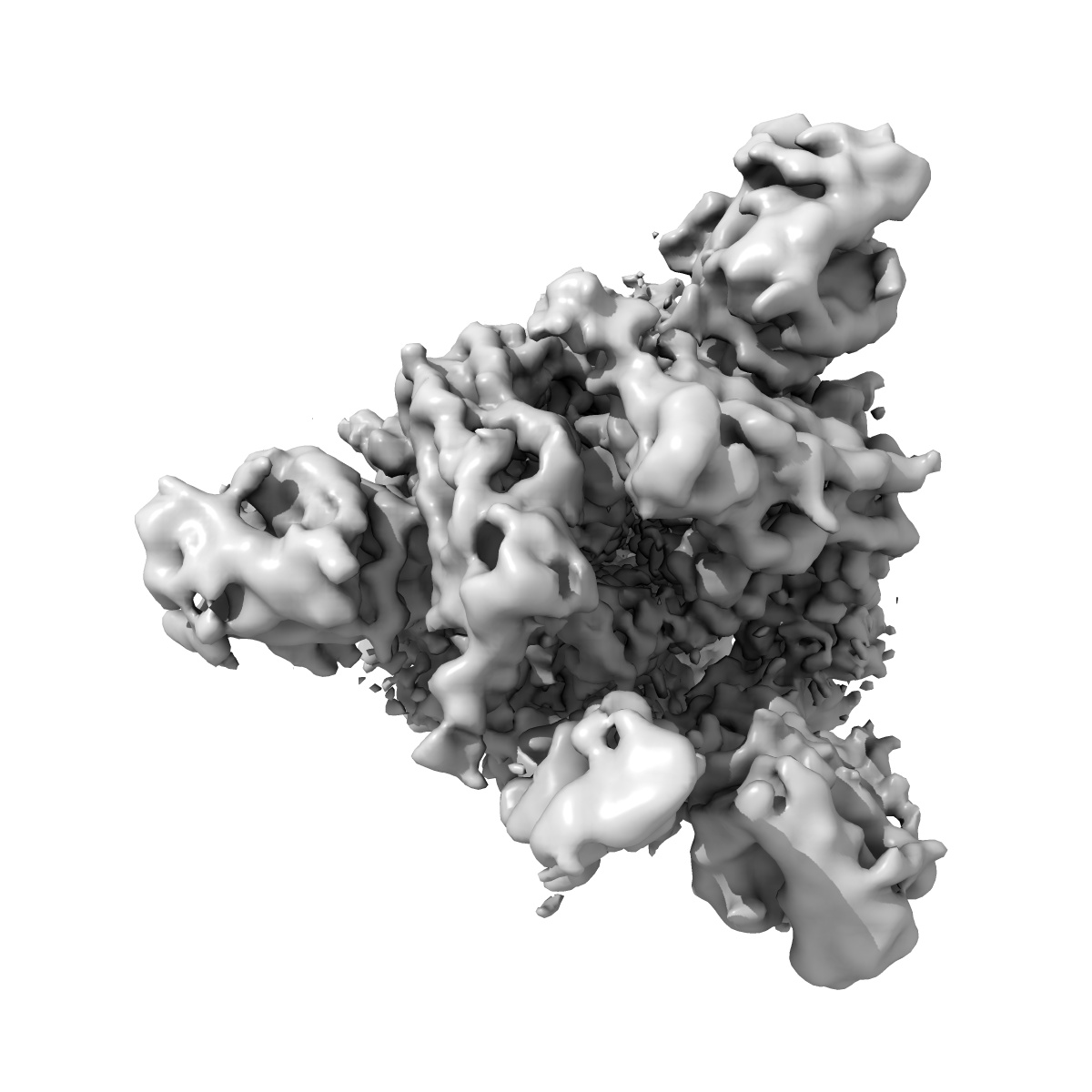
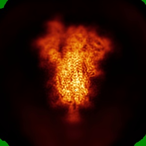
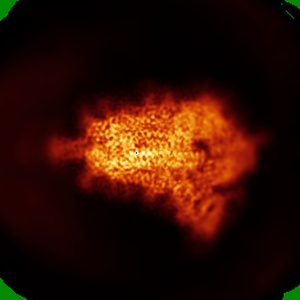
Cryo-EM structure of SARS-CoV-2 Spike Proteins on intact virions: Alpha (B.1.1.7) variant 1 open RBD
EMD-45866
Single-particle4.0 Å
 Deposition: 22/07/2024
Map released: 27/11/2024
Last modified: 25/12/2024
Sample Organism:
Severe acute respiratory syndrome coronavirus 2
Sample: Severe acute respiratory syndrome coronavirus 2
Fitted models: 9crf (Avg. Q-score: 0.349)
Deposition Authors: Ke Z ,
Croll TI ,
Briggs JAG
,
Croll TI ,
Briggs JAG
Sample: Severe acute respiratory syndrome coronavirus 2
Fitted models: 9crf (Avg. Q-score: 0.349)
Deposition Authors: Ke Z
 ,
Croll TI ,
Briggs JAG
,
Croll TI ,
Briggs JAG
Virion morphology and on-virus spike protein structures of diverse SARS-CoV-2 variants.
Ke Z ,
Peacock TP ,
Brown JC ,
Sheppard CM ,
Croll TI ,
Kotecha A ,
Goldhill DH ,
Barclay WS ,
Briggs JAG
(2024) EMBO J , 43 , 6469 - 6495
,
Peacock TP ,
Brown JC ,
Sheppard CM ,
Croll TI ,
Kotecha A ,
Goldhill DH ,
Barclay WS ,
Briggs JAG
(2024) EMBO J , 43 , 6469 - 6495
Abstract:
The evolution of SARS-CoV-2 variants with increased fitness has been accompanied by structural changes in the spike (S) proteins, which are the major target for the adaptive immune response. Single-particle cryo-EM analysis of soluble S protein from SARS-CoV-2 variants has revealed this structural adaptation at high resolution. The analysis of S trimers in situ on intact virions has the potential to provide more functionally relevant insights into S structure and virion morphology. Here, we characterized B.1, Alpha, Beta, Gamma, Delta, Kappa, and Mu variants by cryo-electron microscopy and tomography, assessing S cleavage, virion morphology, S incorporation, "in-situ" high-resolution S structures, and the range of S conformational states. We found no evidence for adaptive changes in virion morphology, but describe multiple different positions in the S protein where amino acid changes alter local protein structure. Taken together, our data are consistent with a model where amino acid changes at multiple positions from the top to the base of the spike cause structural changes that can modulate the conformational dynamics of the S protein.
The evolution of SARS-CoV-2 variants with increased fitness has been accompanied by structural changes in the spike (S) proteins, which are the major target for the adaptive immune response. Single-particle cryo-EM analysis of soluble S protein from SARS-CoV-2 variants has revealed this structural adaptation at high resolution. The analysis of S trimers in situ on intact virions has the potential to provide more functionally relevant insights into S structure and virion morphology. Here, we characterized B.1, Alpha, Beta, Gamma, Delta, Kappa, and Mu variants by cryo-electron microscopy and tomography, assessing S cleavage, virion morphology, S incorporation, "in-situ" high-resolution S structures, and the range of S conformational states. We found no evidence for adaptive changes in virion morphology, but describe multiple different positions in the S protein where amino acid changes alter local protein structure. Taken together, our data are consistent with a model where amino acid changes at multiple positions from the top to the base of the spike cause structural changes that can modulate the conformational dynamics of the S protein.