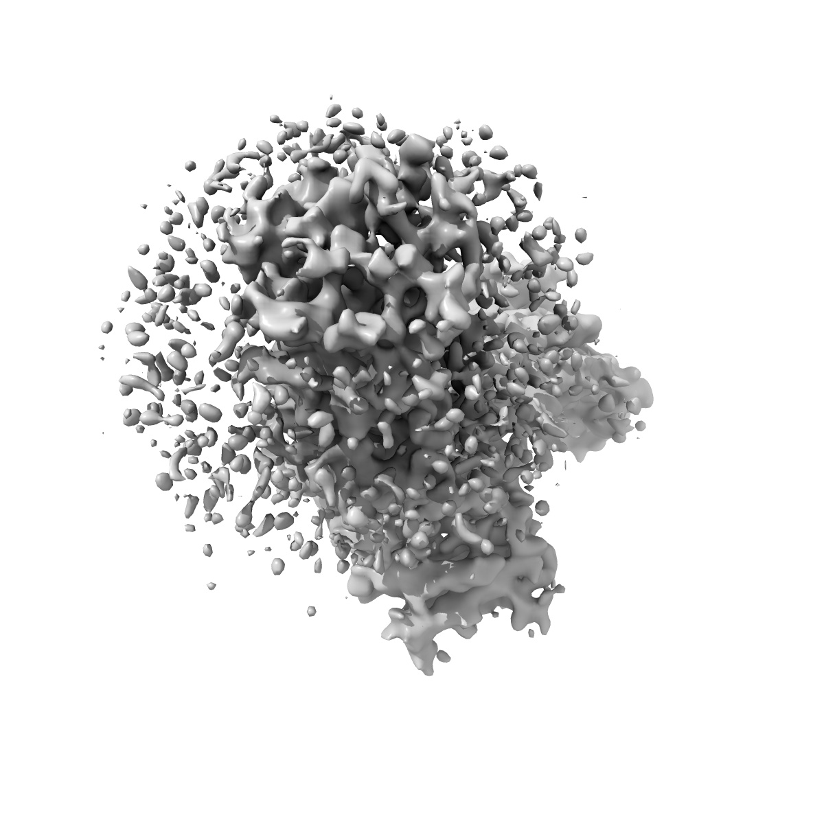
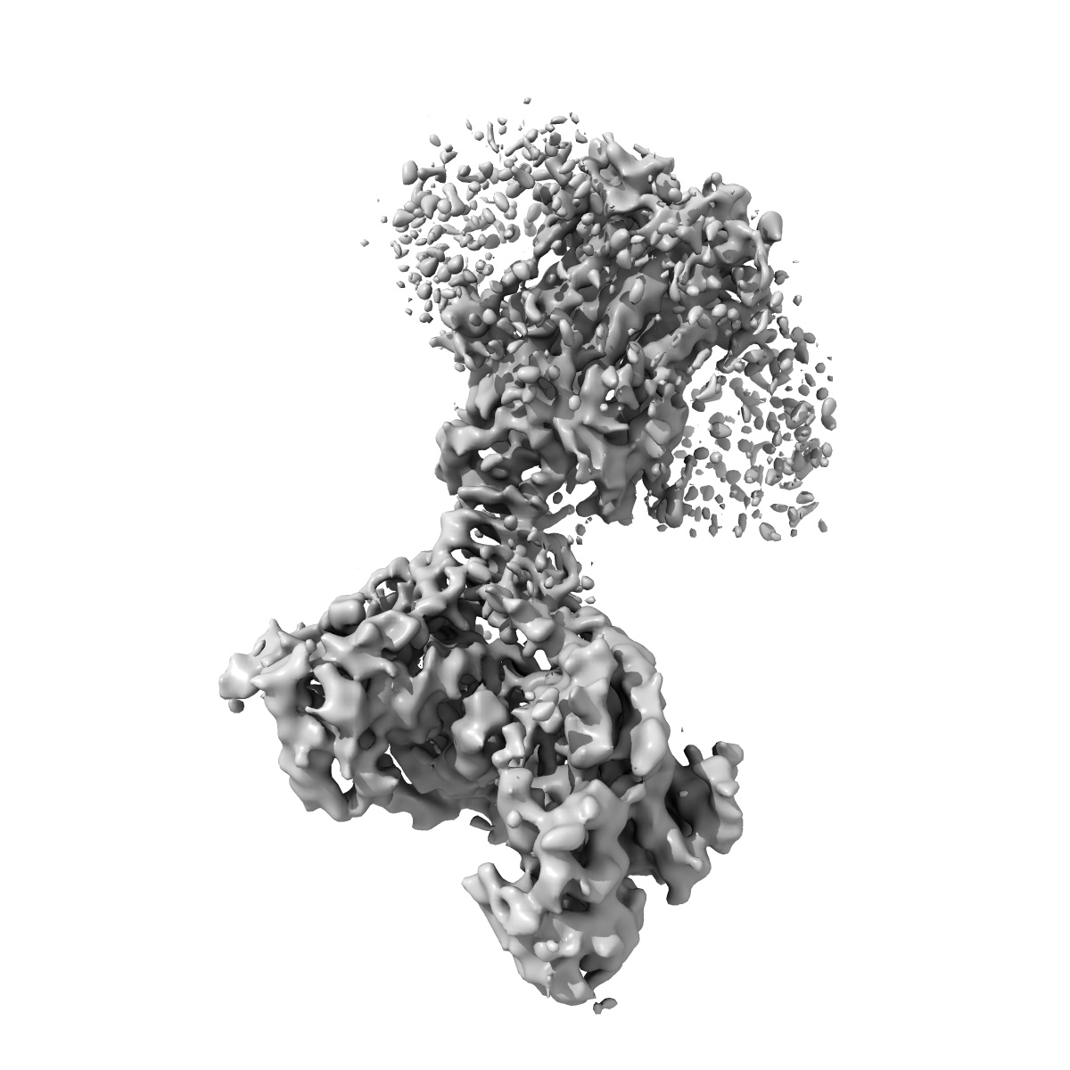
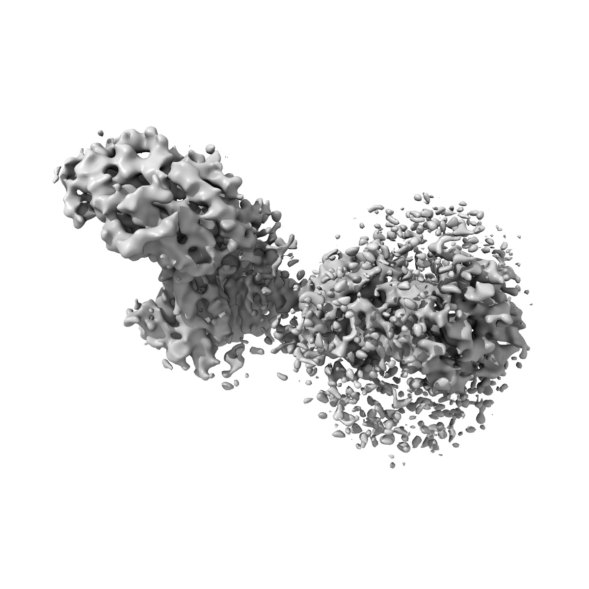
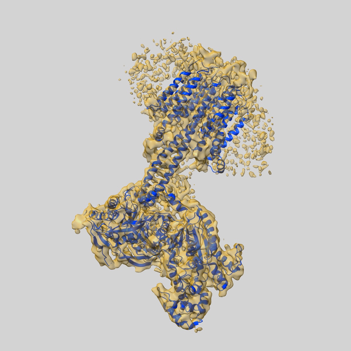
EMD-4721
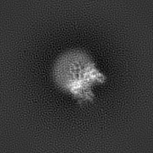
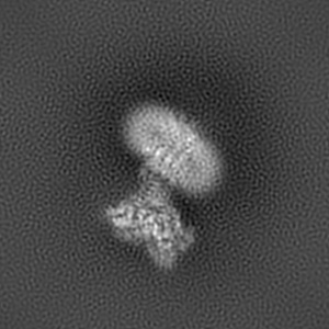
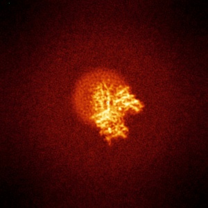
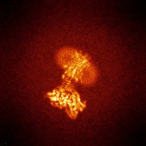
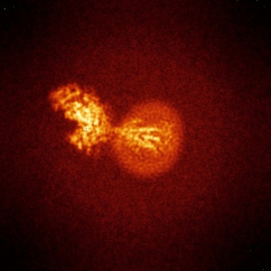
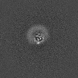
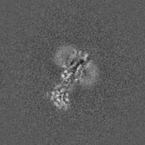
Structure of a truncated adenylyl cyclase bound to MANT-GTP, forskolin and an activatedstimulatory Galphas protein
EMD-4721
Single-particle4.2 Å
 Deposition: 22/03/2019
Map released: 08/05/2019
Last modified: 15/05/2024
Sample Organism:
Bos taurus
Sample: Adenylyl cyclase AC9 (truncation, residues 1-1250) bound to MANT-GTP, forskolin and GalphaS protein
Fitted models: 6r4o (Avg. Q-score: 0.268)
Deposition Authors: Qi C ,
Sorrentino S
,
Sorrentino S
Sample: Adenylyl cyclase AC9 (truncation, residues 1-1250) bound to MANT-GTP, forskolin and GalphaS protein
Fitted models: 6r4o (Avg. Q-score: 0.268)
Deposition Authors: Qi C
 ,
Sorrentino S
,
Sorrentino S
The structure of a membrane adenylyl cyclase bound to an activated stimulatory G protein.
Abstract:
Membrane-integral adenylyl cyclases (ACs) are key enzymes in mammalian heterotrimeric GTP-binding protein (G protein)-dependent signal transduction, which is important in many cellular processes. Signals received by the G protein-coupled receptors are conveyed to ACs through G proteins to modulate the levels of cellular cyclic adenosine monophosphate (cAMP). Here, we describe the cryo-electron microscopy structure of the bovine membrane AC9 bound to an activated G protein αs subunit at 3.4-angstrom resolution. The structure reveals the organization of the membrane domain and helical domain that spans between the membrane and catalytic domains of AC9. The carboxyl-terminal extension of the catalytic domain occludes both the catalytic and the allosteric sites of AC9, inducing a conformation distinct from the substrate- and activator-bound state, suggesting a regulatory role in cAMP production.
Membrane-integral adenylyl cyclases (ACs) are key enzymes in mammalian heterotrimeric GTP-binding protein (G protein)-dependent signal transduction, which is important in many cellular processes. Signals received by the G protein-coupled receptors are conveyed to ACs through G proteins to modulate the levels of cellular cyclic adenosine monophosphate (cAMP). Here, we describe the cryo-electron microscopy structure of the bovine membrane AC9 bound to an activated G protein αs subunit at 3.4-angstrom resolution. The structure reveals the organization of the membrane domain and helical domain that spans between the membrane and catalytic domains of AC9. The carboxyl-terminal extension of the catalytic domain occludes both the catalytic and the allosteric sites of AC9, inducing a conformation distinct from the substrate- and activator-bound state, suggesting a regulatory role in cAMP production.