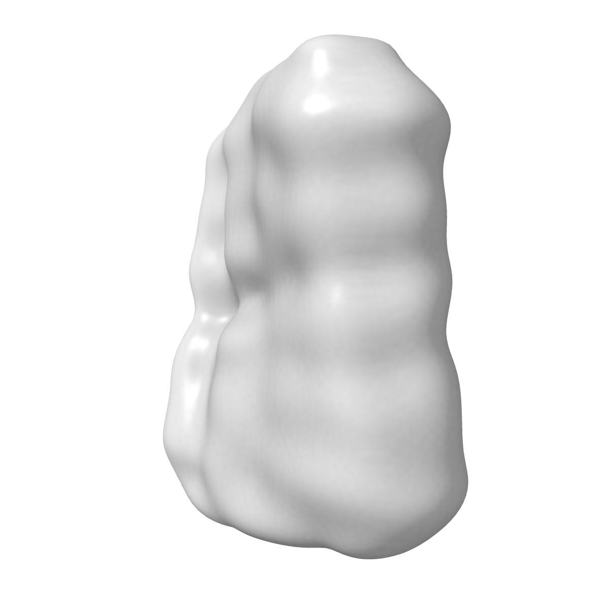
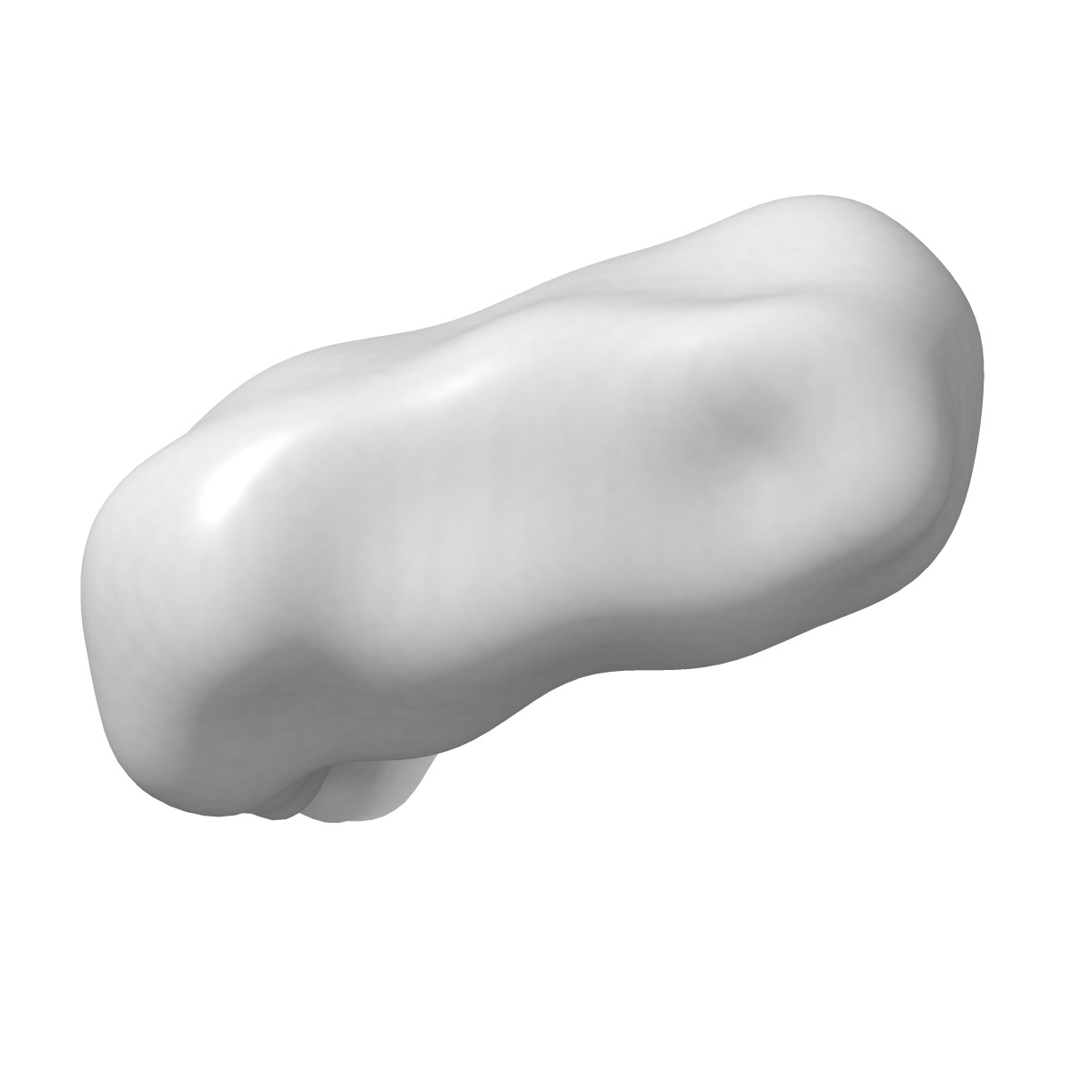
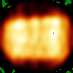
EMD-50155
Tau PHF subtomogram average relating to CS4 extended data Figure 9d
EMD-50155
Subtomogram averaging28.56 Å
 Deposition: 23/04/2024
Map released: 10/07/2024
Last modified: 07/08/2024
Sample Organism:
Homo sapiens
Sample: Microtubule-associated protein tau
Raw data: EMPIAR-12091, EMPIAR-12092, EMPIAR-12088, EMPIAR-12082
Deposition Authors: Jenkins J
Sample: Microtubule-associated protein tau
Raw data: EMPIAR-12091, EMPIAR-12092, EMPIAR-12088, EMPIAR-12082
Deposition Authors: Jenkins J

CryoET of beta-amyloid and tau within postmortem Alzheimer's disease brain.
Gilbert MAG ,
Fatima N,
Jenkins J ,
O'Sullivan TJ,
Schertel A,
Halfon Y,
Wilkinson M ,
Morrema THJ,
Geibel M,
Read RJ ,
Ranson NA ,
Radford SE ,
Hoozemans JJM ,
Frank RAW
(2024) Nature , 631 , 913 - 919
,
Fatima N,
Jenkins J ,
O'Sullivan TJ,
Schertel A,
Halfon Y,
Wilkinson M ,
Morrema THJ,
Geibel M,
Read RJ ,
Ranson NA ,
Radford SE ,
Hoozemans JJM ,
Frank RAW
(2024) Nature , 631 , 913 - 919
Abstract:
A defining pathological feature of most neurodegenerative diseases is the assembly of proteins into amyloid that form disease-specific structures1. In Alzheimer's disease, this is characterized by the deposition of β-amyloid and tau with disease-specific conformations. The in situ structure of amyloid in the human brain is unknown. Here, using cryo-fluorescence microscopy-targeted cryo-sectioning, cryo-focused ion beam-scanning electron microscopy lift-out and cryo-electron tomography, we determined in-tissue architectures of β-amyloid and tau pathology in a postmortem Alzheimer's disease donor brain. β-amyloid plaques contained a mixture of fibrils, some of which were branched, and protofilaments, arranged in parallel arrays and lattice-like structures. Extracellular vesicles and cuboidal particles defined the non-amyloid constituents of β-amyloid plaques. By contrast, tau inclusions formed parallel clusters of unbranched filaments. Subtomogram averaging a cluster of 136 tau filaments in a single tomogram revealed the polypeptide backbone conformation and filament polarity orientation of paired helical filaments within tissue. Filaments within most clusters were similar to each other, but were different between clusters, showing amyloid heterogeneity that is spatially organized by subcellular location. The in situ structural approaches outlined here for human donor tissues have applications to a broad range of neurodegenerative diseases.
A defining pathological feature of most neurodegenerative diseases is the assembly of proteins into amyloid that form disease-specific structures1. In Alzheimer's disease, this is characterized by the deposition of β-amyloid and tau with disease-specific conformations. The in situ structure of amyloid in the human brain is unknown. Here, using cryo-fluorescence microscopy-targeted cryo-sectioning, cryo-focused ion beam-scanning electron microscopy lift-out and cryo-electron tomography, we determined in-tissue architectures of β-amyloid and tau pathology in a postmortem Alzheimer's disease donor brain. β-amyloid plaques contained a mixture of fibrils, some of which were branched, and protofilaments, arranged in parallel arrays and lattice-like structures. Extracellular vesicles and cuboidal particles defined the non-amyloid constituents of β-amyloid plaques. By contrast, tau inclusions formed parallel clusters of unbranched filaments. Subtomogram averaging a cluster of 136 tau filaments in a single tomogram revealed the polypeptide backbone conformation and filament polarity orientation of paired helical filaments within tissue. Filaments within most clusters were similar to each other, but were different between clusters, showing amyloid heterogeneity that is spatially organized by subcellular location. The in situ structural approaches outlined here for human donor tissues have applications to a broad range of neurodegenerative diseases.