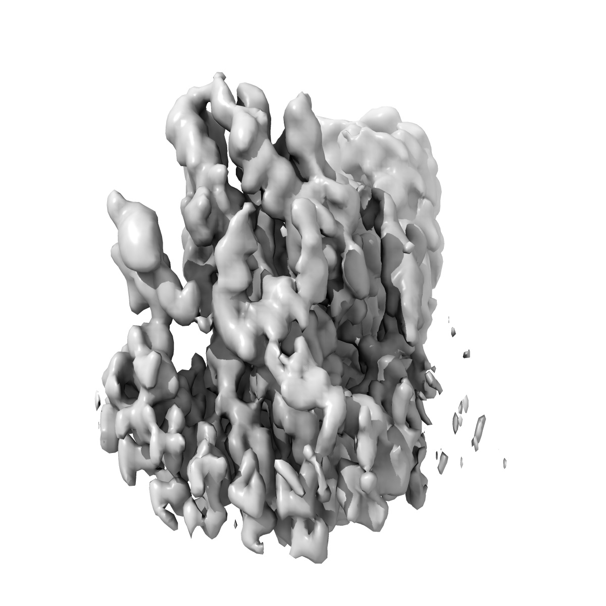
EMD-51467
Focussed refinement on alpha-Latrotoxin, ChainA, residues 1-795
EMD-51467
Single-particle2.75 Å
 Deposition: 02/09/2024
Map released: 16/10/2024
Last modified: 16/10/2024
Sample Organism:
Latrodectus tredecimguttatus
Sample: tetrameric complex of alpha-latrotoxin
Deposition Authors: Klink BU ,
Gatsogiannis C ,
Kalyankumar KS
,
Gatsogiannis C ,
Kalyankumar KS
Sample: tetrameric complex of alpha-latrotoxin
Deposition Authors: Klink BU
 ,
Gatsogiannis C ,
Kalyankumar KS
,
Gatsogiannis C ,
Kalyankumar KS
Structural basis of alpha-latrotoxin transition to a cation-selective pore.
Klink BU ,
Alavizargar A ,
Kalyankumar KS ,
Chen M ,
Heuer A ,
Gatsogiannis C
(2024) Nat Commun , 15 , 8551 - 8551
,
Alavizargar A ,
Kalyankumar KS ,
Chen M ,
Heuer A ,
Gatsogiannis C
(2024) Nat Commun , 15 , 8551 - 8551
Abstract:
The potent neurotoxic venom of the black widow spider contains a cocktail of seven phylum-specific latrotoxins (LTXs), but only one, α-LTX, targets vertebrates. This 130 kDa toxin binds to receptors at presynaptic nerve terminals and triggers a massive release of neurotransmitters. It is widely accepted that LTXs tetramerize and insert into the presynaptic membrane, thereby forming Ca2+-conductive pores, but the underlying mechanism remains poorly understood. LTXs are homologous and consist of an N-terminal region with three distinct domains, along with a C-terminal domain containing up to 22 consecutive ankyrin repeats. Here we report cryoEM structures of the vertebrate-specific α-LTX tetramer in its prepore and pore state. Our structures, in combination with AlphaFold2-based structural modeling and molecular dynamics simulations, reveal dramatic conformational changes in the N-terminal region of the complex. Four distinct helical bundles rearrange and together form a highly stable, 15 nm long, cation-impermeable coiled-coil stalk. This stalk, in turn, positions an N-terminal pair of helices within the membrane, thereby enabling the assembly of a cation-permeable channel. Taken together, these data give insight into a unique mechanism for membrane insertion and channel formation, characteristic of the LTX family, and provide the necessary framework for advancing novel therapeutics and biotechnological applications.
The potent neurotoxic venom of the black widow spider contains a cocktail of seven phylum-specific latrotoxins (LTXs), but only one, α-LTX, targets vertebrates. This 130 kDa toxin binds to receptors at presynaptic nerve terminals and triggers a massive release of neurotransmitters. It is widely accepted that LTXs tetramerize and insert into the presynaptic membrane, thereby forming Ca2+-conductive pores, but the underlying mechanism remains poorly understood. LTXs are homologous and consist of an N-terminal region with three distinct domains, along with a C-terminal domain containing up to 22 consecutive ankyrin repeats. Here we report cryoEM structures of the vertebrate-specific α-LTX tetramer in its prepore and pore state. Our structures, in combination with AlphaFold2-based structural modeling and molecular dynamics simulations, reveal dramatic conformational changes in the N-terminal region of the complex. Four distinct helical bundles rearrange and together form a highly stable, 15 nm long, cation-impermeable coiled-coil stalk. This stalk, in turn, positions an N-terminal pair of helices within the membrane, thereby enabling the assembly of a cation-permeable channel. Taken together, these data give insight into a unique mechanism for membrane insertion and channel formation, characteristic of the LTX family, and provide the necessary framework for advancing novel therapeutics and biotechnological applications.