EMD-51627
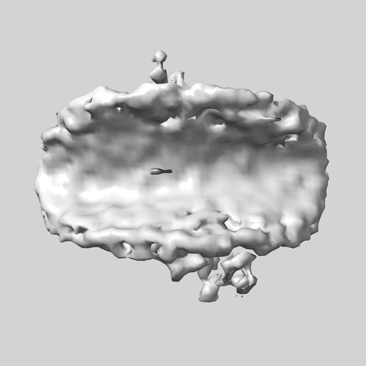
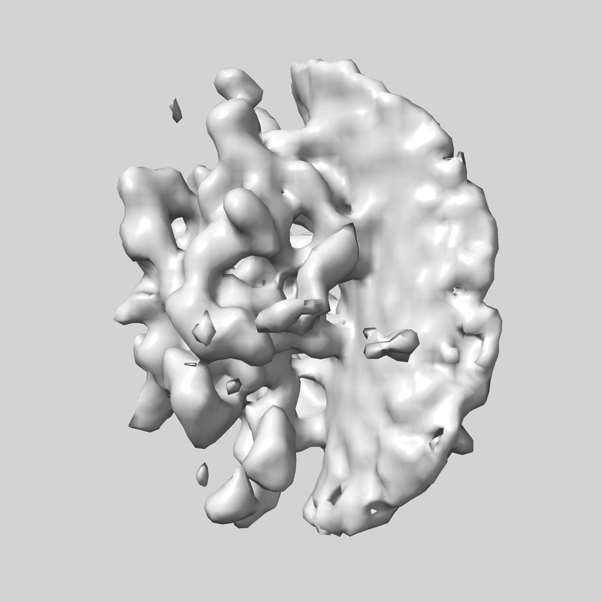
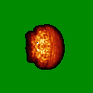
In situ human inner ring subunit of nuclear pore complex (FIB-lamella data of HIV infected macrophages)
EMD-51627
Subtomogram averaging28.5 Å
 Deposition: 23/09/2024
Map released: 15/01/2025
Last modified: 05/02/2025
Sample Organism:
Homo sapiens
Sample: In situ human inner ring subunit of nuclear pore complex (FIB-lamella data of HIV infected macrophages)
Deposition Authors: Kreysing JP ,
Welsch S ,
Turonova B ,
Beck M
,
Welsch S ,
Turonova B ,
Beck M
Sample: In situ human inner ring subunit of nuclear pore complex (FIB-lamella data of HIV infected macrophages)
Deposition Authors: Kreysing JP
 ,
Welsch S ,
Turonova B ,
Beck M
,
Welsch S ,
Turonova B ,
Beck M
Passage of the HIV capsid cracks the nuclear pore.
Kreysing JP ,
Heidari M ,
Zila V ,
Cruz-Leon S ,
Obarska-Kosinska A ,
Laketa V ,
Rohleder L ,
Welsch S ,
Kofinger J,
Turonova B ,
Hummer G ,
Krausslich HG,
Beck M
(2025) Cell
,
Heidari M ,
Zila V ,
Cruz-Leon S ,
Obarska-Kosinska A ,
Laketa V ,
Rohleder L ,
Welsch S ,
Kofinger J,
Turonova B ,
Hummer G ,
Krausslich HG,
Beck M
(2025) Cell
Abstract:
Upon infection, human immunodeficiency virus type 1 (HIV-1) releases its cone-shaped capsid into the cytoplasm of infected T cells and macrophages. The capsid enters the nuclear pore complex (NPC), driven by interactions with numerous phenylalanine-glycine (FG)-repeat nucleoporins (FG-Nups). Whether NPCs structurally adapt to capsid passage and whether capsids are modified during passage remains unknown, however. Here, we combined super-resolution and correlative microscopy with cryoelectron tomography and molecular simulations to study the nuclear entry of HIV-1 capsids in primary human macrophages. Our data indicate that cytosolically bound cyclophilin A is stripped off capsids entering the NPC, and the capsid hexagonal lattice remains largely intact inside and beyond the central channel. Strikingly, the NPC scaffold rings frequently crack during capsid passage, consistent with computer simulations indicating the need for NPC widening. The unique cone shape of the HIV-1 capsid facilitates its entry into NPCs and helps to crack their rings.
Upon infection, human immunodeficiency virus type 1 (HIV-1) releases its cone-shaped capsid into the cytoplasm of infected T cells and macrophages. The capsid enters the nuclear pore complex (NPC), driven by interactions with numerous phenylalanine-glycine (FG)-repeat nucleoporins (FG-Nups). Whether NPCs structurally adapt to capsid passage and whether capsids are modified during passage remains unknown, however. Here, we combined super-resolution and correlative microscopy with cryoelectron tomography and molecular simulations to study the nuclear entry of HIV-1 capsids in primary human macrophages. Our data indicate that cytosolically bound cyclophilin A is stripped off capsids entering the NPC, and the capsid hexagonal lattice remains largely intact inside and beyond the central channel. Strikingly, the NPC scaffold rings frequently crack during capsid passage, consistent with computer simulations indicating the need for NPC widening. The unique cone shape of the HIV-1 capsid facilitates its entry into NPCs and helps to crack their rings.