EMD-5844
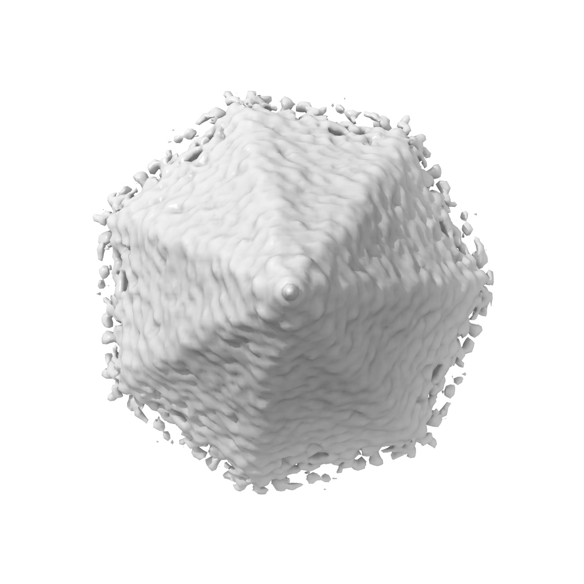
Subtomogram average of a virus-associated pyramid
EMD-5844
Subtomogram averaging Deposition: 19/12/2013
Map released: 19/02/2014
Last modified: 23/04/2014
Sample Organism:
Sulfolobus islandicus rod-shaped virus 2
Sample: Reconstruction of a virus-associated pyramid induced by overexpressing the viral protein PVAP in E. coli
Deposition Authors: Daum B ,
Kuehlbrandt W
,
Kuehlbrandt W
Sample: Reconstruction of a virus-associated pyramid induced by overexpressing the viral protein PVAP in E. coli
Deposition Authors: Daum B
 ,
Kuehlbrandt W
,
Kuehlbrandt W
Self-assembly of the general membrane-remodeling protein PVAP into sevenfold virus-associated pyramids.
Daum B ,
Quax TE ,
Sachse M ,
Mills DJ ,
Reimann J,
Yildiz O ,
Hader S,
Saveanu C ,
Forterre P,
Albers SV ,
Kuhlbrandt W ,
Prangishvili D
(2014) PNAS , 111 , 3829 - 3834
,
Quax TE ,
Sachse M ,
Mills DJ ,
Reimann J,
Yildiz O ,
Hader S,
Saveanu C ,
Forterre P,
Albers SV ,
Kuhlbrandt W ,
Prangishvili D
(2014) PNAS , 111 , 3829 - 3834
Abstract:
Viruses have developed a wide range of strategies to escape from the host cells in which they replicate. For egress some archaeal viruses use a pyramidal structure with sevenfold rotational symmetry. Virus-associated pyramids (VAPs) assemble in the host cell membrane from the virus-encoded protein PVAP and open at the end of the infection cycle. We characterize this unusual supramolecular assembly using a combination of genetic, biochemical, and electron microscopic techniques. By whole-cell electron cryotomography, we monitored morphological changes in virus-infected host cells. Subtomogram averaging reveals the VAP structure. By heterologous expression of PVAP in cells from all three domains of life, we demonstrate that the protein integrates indiscriminately into virtually any biological membrane, where it forms sevenfold pyramids. We identify the protein domains essential for VAP formation in PVAP truncation mutants by their ability to remodel the cell membrane. Self-assembly of PVAP into pyramids requires at least two different, in-plane and out-of-plane, protein interactions. Our findings allow us to propose a model describing how PVAP arranges to form sevenfold pyramids and suggest how this small, robust protein may be used as a general membrane-remodeling system.
Viruses have developed a wide range of strategies to escape from the host cells in which they replicate. For egress some archaeal viruses use a pyramidal structure with sevenfold rotational symmetry. Virus-associated pyramids (VAPs) assemble in the host cell membrane from the virus-encoded protein PVAP and open at the end of the infection cycle. We characterize this unusual supramolecular assembly using a combination of genetic, biochemical, and electron microscopic techniques. By whole-cell electron cryotomography, we monitored morphological changes in virus-infected host cells. Subtomogram averaging reveals the VAP structure. By heterologous expression of PVAP in cells from all three domains of life, we demonstrate that the protein integrates indiscriminately into virtually any biological membrane, where it forms sevenfold pyramids. We identify the protein domains essential for VAP formation in PVAP truncation mutants by their ability to remodel the cell membrane. Self-assembly of PVAP into pyramids requires at least two different, in-plane and out-of-plane, protein interactions. Our findings allow us to propose a model describing how PVAP arranges to form sevenfold pyramids and suggest how this small, robust protein may be used as a general membrane-remodeling system.