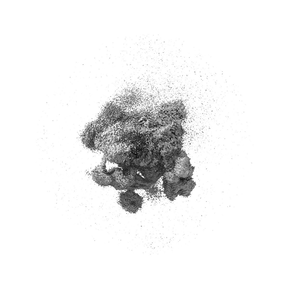
EMD-6415
Local map for reduced Spp42 region of the yeast spliceosome at 3.52 angstrom resolution
EMD-6415
Single-particle3.52 Å
 Deposition: 09/08/2015
Map released: 16/09/2015
Last modified: 08/06/2016
Sample Organism:
Schizosaccharomyces pombe
Sample: the yeast spliceosome
Fitted models: 3jb9 (Avg. Q-score: 0.127)
Deposition Authors: Yan C ,
Hang J,
Wan R,
Huang M,
Wong C,
Shi Y
,
Hang J,
Wan R,
Huang M,
Wong C,
Shi Y
Sample: the yeast spliceosome
Fitted models: 3jb9 (Avg. Q-score: 0.127)
Deposition Authors: Yan C
 ,
Hang J,
Wan R,
Huang M,
Wong C,
Shi Y
,
Hang J,
Wan R,
Huang M,
Wong C,
Shi Y
Structure of a yeast spliceosome at 3.6-angstrom resolution
Abstract:
Splicing of precursor messenger RNA (pre-mRNA) in yeast is executed by the spliceosome, which consists of five small nuclear ribonucleoproteins (snRNPs), NTC (nineteen complex), NTC-related proteins (NTR), and a number of associated enzymes and cofactors. Here, we report the three-dimensional structure of a Schizosaccharomyces pombe spliceosome at 3.6-angstrom resolution, revealed by means of single-particle cryogenic electron microscopy. This spliceosome contains U2 and U5 snRNPs, NTC, NTR, U6 small nuclear RNA, and an RNA intron lariat. The atomic model includes 10,574 amino acids from 37 proteins and four RNA molecules, with a combined molecular mass of approximately 1.3 megadaltons. Spp42 (Prp8 in Saccharomyces cerevisiae), the key protein component of the U5 snRNP, forms a central scaffold and anchors the catalytic center. Both the morphology and the placement of protein components appear to have evolved to facilitate the dynamic process of pre-mRNA splicing. Our near-atomic-resolution structure of a central spliceosome provides a molecular framework for mechanistic understanding of pre-mRNA splicing.
Splicing of precursor messenger RNA (pre-mRNA) in yeast is executed by the spliceosome, which consists of five small nuclear ribonucleoproteins (snRNPs), NTC (nineteen complex), NTC-related proteins (NTR), and a number of associated enzymes and cofactors. Here, we report the three-dimensional structure of a Schizosaccharomyces pombe spliceosome at 3.6-angstrom resolution, revealed by means of single-particle cryogenic electron microscopy. This spliceosome contains U2 and U5 snRNPs, NTC, NTR, U6 small nuclear RNA, and an RNA intron lariat. The atomic model includes 10,574 amino acids from 37 proteins and four RNA molecules, with a combined molecular mass of approximately 1.3 megadaltons. Spp42 (Prp8 in Saccharomyces cerevisiae), the key protein component of the U5 snRNP, forms a central scaffold and anchors the catalytic center. Both the morphology and the placement of protein components appear to have evolved to facilitate the dynamic process of pre-mRNA splicing. Our near-atomic-resolution structure of a central spliceosome provides a molecular framework for mechanistic understanding of pre-mRNA splicing.