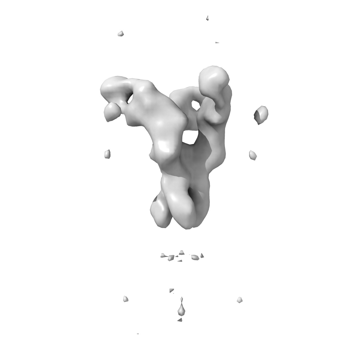
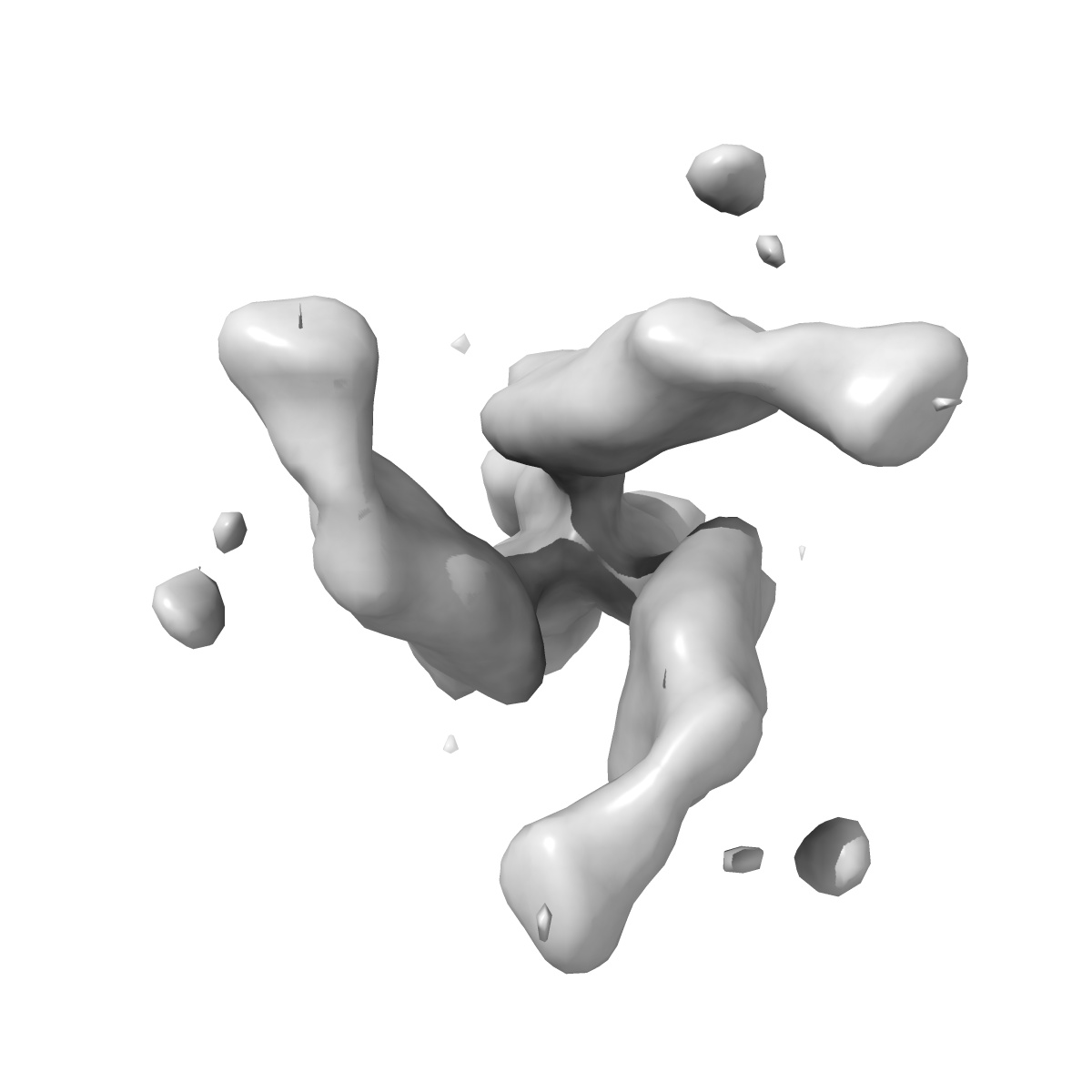
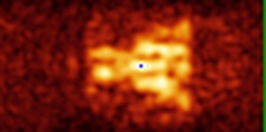
EMD-6613
Cryo-electron microscopy structure of chimeric influenza cH5/1 hemagglutinin bound to head-targeting antibody, 3F5
EMD-6613
Subtomogram averaging25.0 Å
 Deposition: 29/02/2016
Map released: 30/03/2016
Last modified: 13/04/2016
Sample Organism:
unidentified influenza virus,
Mus musculus
Sample: Tomographic subvolume average of virus-bound influenza cH5/1 HA in complex with the head-binding antibody, 3F5
Deposition Authors: Tran EEH, Podolsky KA ,
Bartesaghi A,
Kuybeda O,
Grandinetti G,
Wohlbold TJ,
Tan GS ,
Nachbagauer R ,
Palese P ,
Krammer F ,
Subramaniam S
,
Bartesaghi A,
Kuybeda O,
Grandinetti G,
Wohlbold TJ,
Tan GS ,
Nachbagauer R ,
Palese P ,
Krammer F ,
Subramaniam S
Sample: Tomographic subvolume average of virus-bound influenza cH5/1 HA in complex with the head-binding antibody, 3F5
Deposition Authors: Tran EEH, Podolsky KA
 ,
Bartesaghi A,
Kuybeda O,
Grandinetti G,
Wohlbold TJ,
Tan GS ,
Nachbagauer R ,
Palese P ,
Krammer F ,
Subramaniam S
,
Bartesaghi A,
Kuybeda O,
Grandinetti G,
Wohlbold TJ,
Tan GS ,
Nachbagauer R ,
Palese P ,
Krammer F ,
Subramaniam S
Cryo-electron Microscopy Structures of Chimeric Hemagglutinin Displayed on a Universal Vaccine Candidate
Tran EEH,
Podolsky KA ,
Bartesaghi A,
Kuybeda O,
Grandinetti G,
Wohlbold TJ,
Tan GS ,
Nachbagauer R ,
Palese P ,
Krammer F ,
Subramaniam S
(2016) mBio , 7
,
Bartesaghi A,
Kuybeda O,
Grandinetti G,
Wohlbold TJ,
Tan GS ,
Nachbagauer R ,
Palese P ,
Krammer F ,
Subramaniam S
(2016) mBio , 7
Abstract: