EMD-6770
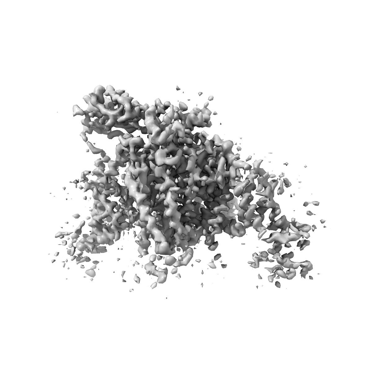
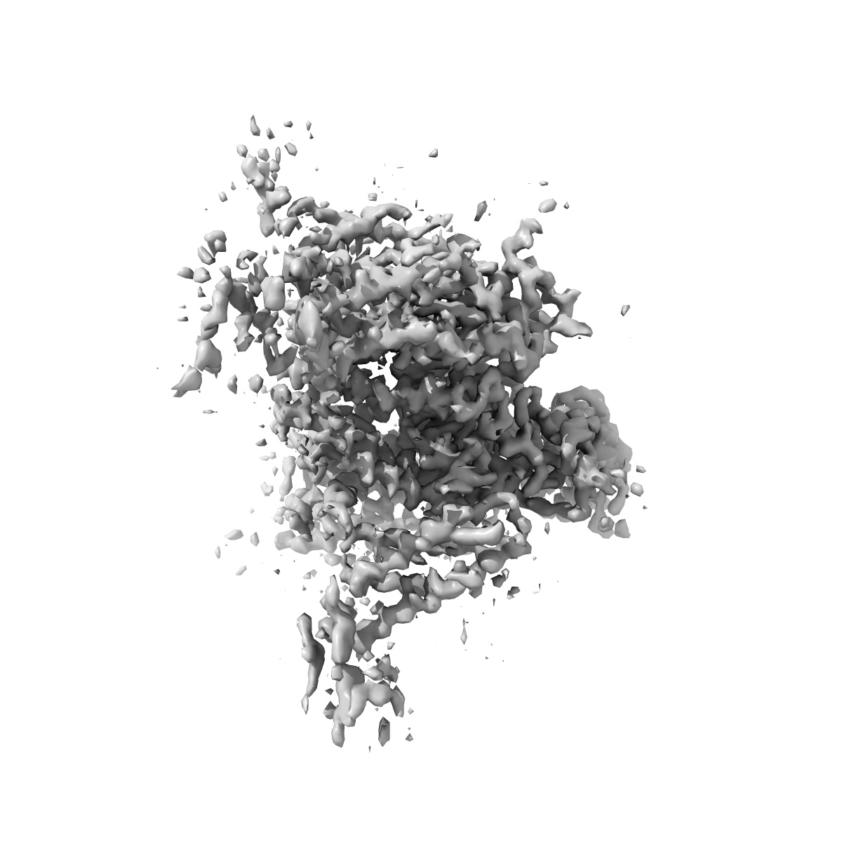
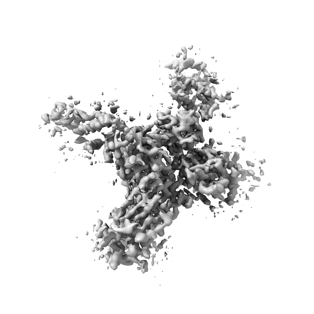
Structure of the Nav1.4-beta1 complex from electric eel
EMD-6770
Single-particle4.0 Å
 Deposition: 15/06/2017
Map released: 09/08/2017
Last modified: 30/10/2024
Sample Organism:
Electrophorus electricus
Sample: voltage gated sodium channel EeNav1.4
Fitted models: 5xsy (Avg. Q-score: 0.403)
Deposition Authors: Yan Z, Zhou Q
Sample: voltage gated sodium channel EeNav1.4
Fitted models: 5xsy (Avg. Q-score: 0.403)
Deposition Authors: Yan Z, Zhou Q
Structure of the Nav1.4-beta 1 Complex from Electric Eel.
Yan Z,
Zhou Q,
Wang L,
Wu J  ,
Zhao Y,
Huang G,
Peng W ,
Shen H,
Lei J,
Yan N
,
Zhao Y,
Huang G,
Peng W ,
Shen H,
Lei J,
Yan N
(2017) Cell , 170 , 470 - 482.e11
 ,
Zhao Y,
Huang G,
Peng W ,
Shen H,
Lei J,
Yan N
,
Zhao Y,
Huang G,
Peng W ,
Shen H,
Lei J,
Yan N
(2017) Cell , 170 , 470 - 482.e11
Abstract:
Voltage-gated sodium (Nav) channels initiate and propagate action potentials. Here, we present the cryo-EM structure of EeNav1.4, the Nav channel from electric eel, in complex with the β1 subunit at 4.0 Å resolution. The immunoglobulin domain of β1 docks onto the extracellular L5I and L6IV loops of EeNav1.4 via extensive polar interactions, and the single transmembrane helix interacts with the third voltage-sensing domain (VSDIII). The VSDs exhibit "up" conformations, while the intracellular gate of the pore domain is kept open by a digitonin-like molecule. Structural comparison with closed NavPaS shows that the outward transfer of gating charges is coupled to the iris-like pore domain dilation through intricate force transmissions involving multiple channel segments. The IFM fast inactivation motif on the III-IV linker is plugged into the corner enclosed by the outer S4-S5 and inner S6 segments in repeats III and IV, suggesting a potential allosteric blocking mechanism for fast inactivation.
Voltage-gated sodium (Nav) channels initiate and propagate action potentials. Here, we present the cryo-EM structure of EeNav1.4, the Nav channel from electric eel, in complex with the β1 subunit at 4.0 Å resolution. The immunoglobulin domain of β1 docks onto the extracellular L5I and L6IV loops of EeNav1.4 via extensive polar interactions, and the single transmembrane helix interacts with the third voltage-sensing domain (VSDIII). The VSDs exhibit "up" conformations, while the intracellular gate of the pore domain is kept open by a digitonin-like molecule. Structural comparison with closed NavPaS shows that the outward transfer of gating charges is coupled to the iris-like pore domain dilation through intricate force transmissions involving multiple channel segments. The IFM fast inactivation motif on the III-IV linker is plugged into the corner enclosed by the outer S4-S5 and inner S6 segments in repeats III and IV, suggesting a potential allosteric blocking mechanism for fast inactivation.