EMD-7140
Protein in nanodisc on a gold nanowire grid plunged with Spotiton
EMD-7140
Tomography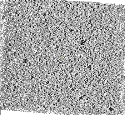 Deposition: 05/12/2017
Map released: 20/12/2017
Last modified: 29/01/2020
Sample Organism:
unidentified
Sample: Unidentified protein in nanodisc
Deposition Authors: Noble AN, Dandey VP, Wei H, Brasch J, Chase J, Acharya P, Tan Y, Zhang Z, Kim LY, Scapin G, Rapp M, Eng ET, Rice WJ, Cheng A, Negro CJ, Shapiro L, Kwong PD, Jeruzalmi D, des Georges A, Potter CS, Carragher B
Sample: Unidentified protein in nanodisc
Deposition Authors: Noble AN, Dandey VP, Wei H, Brasch J, Chase J, Acharya P, Tan Y, Zhang Z, Kim LY, Scapin G, Rapp M, Eng ET, Rice WJ, Cheng A, Negro CJ, Shapiro L, Kwong PD, Jeruzalmi D, des Georges A, Potter CS, Carragher B
Routine single particle CryoEM sample and grid characterization by tomography.
Noble AJ  ,
Dandey VP,
Wei H,
Brasch J,
Chase J,
Acharya P ,
Tan YZ ,
Zhang Z,
Kim LY,
Scapin G,
Rapp M,
Eng ET ,
Rice WJ ,
Cheng A,
Negro CJ,
Shapiro L,
Kwong PD,
Jeruzalmi D ,
des Georges A ,
Potter CS ,
Carragher B
,
Dandey VP,
Wei H,
Brasch J,
Chase J,
Acharya P ,
Tan YZ ,
Zhang Z,
Kim LY,
Scapin G,
Rapp M,
Eng ET ,
Rice WJ ,
Cheng A,
Negro CJ,
Shapiro L,
Kwong PD,
Jeruzalmi D ,
des Georges A ,
Potter CS ,
Carragher B
(2018) eLife , 7
 ,
Dandey VP,
Wei H,
Brasch J,
Chase J,
Acharya P ,
Tan YZ ,
Zhang Z,
Kim LY,
Scapin G,
Rapp M,
Eng ET ,
Rice WJ ,
Cheng A,
Negro CJ,
Shapiro L,
Kwong PD,
Jeruzalmi D ,
des Georges A ,
Potter CS ,
Carragher B
,
Dandey VP,
Wei H,
Brasch J,
Chase J,
Acharya P ,
Tan YZ ,
Zhang Z,
Kim LY,
Scapin G,
Rapp M,
Eng ET ,
Rice WJ ,
Cheng A,
Negro CJ,
Shapiro L,
Kwong PD,
Jeruzalmi D ,
des Georges A ,
Potter CS ,
Carragher B
(2018) eLife , 7
Abstract:
Single particle cryo-electron microscopy (cryoEM) is often performed under the assumption that particles are not adsorbed to the air-water interfaces and in thin, vitreous ice. In this study, we performed fiducial-less tomography on over 50 different cryoEM grid/sample preparations to determine the particle distribution within the ice and the overall geometry of the ice in grid holes. Surprisingly, by studying particles in holes in 3D from over 1000 tomograms, we have determined that the vast majority of particles (approximately 90%) are adsorbed to an air-water interface. The implications of this observation are wide-ranging, with potential ramifications regarding protein denaturation, conformational change, and preferred orientation. We also show that fiducial-less cryo-electron tomography on single particle grids may be used to determine ice thickness, optimal single particle collection areas and strategies, particle heterogeneity, and de novo models for template picking and single particle alignment.
Single particle cryo-electron microscopy (cryoEM) is often performed under the assumption that particles are not adsorbed to the air-water interfaces and in thin, vitreous ice. In this study, we performed fiducial-less tomography on over 50 different cryoEM grid/sample preparations to determine the particle distribution within the ice and the overall geometry of the ice in grid holes. Surprisingly, by studying particles in holes in 3D from over 1000 tomograms, we have determined that the vast majority of particles (approximately 90%) are adsorbed to an air-water interface. The implications of this observation are wide-ranging, with potential ramifications regarding protein denaturation, conformational change, and preferred orientation. We also show that fiducial-less cryo-electron tomography on single particle grids may be used to determine ice thickness, optimal single particle collection areas and strategies, particle heterogeneity, and de novo models for template picking and single particle alignment.