EMD-7540
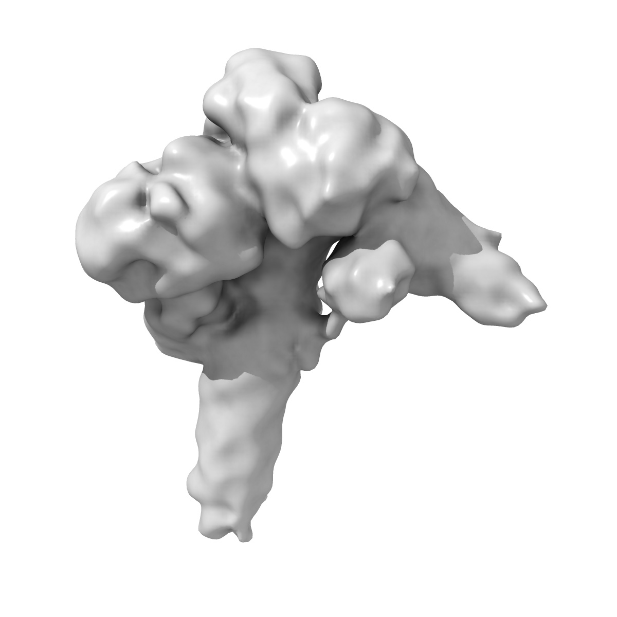
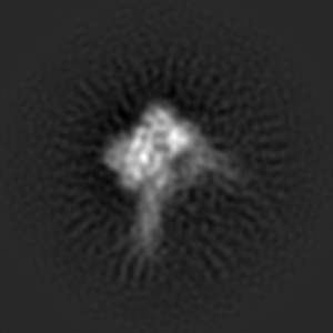
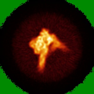
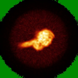
Cryo-EM map of an HIV-1 reverse transcriptase initiation complex in magnesium chloride imaging buffer
EMD-7540
Single-particle8.2 Å
 Deposition: 09/03/2018
Map released: 02/05/2018
Last modified: 09/05/2018
Sample Organism:
Human immunodeficiency virus 1,
Human
Sample: HIV-1 reverse transcriptase initiation complex
Deposition Authors: Larsen KP, Chen DH, Puglisi JD, Puglisi EV
Sample: HIV-1 reverse transcriptase initiation complex
Deposition Authors: Larsen KP, Chen DH, Puglisi JD, Puglisi EV
Architecture of an HIV-1 reverse transcriptase initiation complex.
Larsen KP  ,
Mathiharan YK ,
Kappel K ,
Coey AT,
Chen DH,
Barrero D,
Madigan L,
Puglisi JD,
Skiniotis G ,
Puglisi EV
,
Mathiharan YK ,
Kappel K ,
Coey AT,
Chen DH,
Barrero D,
Madigan L,
Puglisi JD,
Skiniotis G ,
Puglisi EV
(2018) Nature , 557 , 118 - 122
 ,
Mathiharan YK ,
Kappel K ,
Coey AT,
Chen DH,
Barrero D,
Madigan L,
Puglisi JD,
Skiniotis G ,
Puglisi EV
,
Mathiharan YK ,
Kappel K ,
Coey AT,
Chen DH,
Barrero D,
Madigan L,
Puglisi JD,
Skiniotis G ,
Puglisi EV
(2018) Nature , 557 , 118 - 122
Abstract:
Reverse transcription of the HIV-1 RNA genome into double-stranded DNA is a central step in viral infection 1 and a common target of antiretroviral drugs 2 . The reaction is catalysed by viral reverse transcriptase (RT)3,4 that is packaged in an infectious virion with two copies of viral genomic RNA 5 each bound to host lysine 3 transfer RNA (tRNALys3), which acts as a primer for initiation of reverse transcription6,7. Upon viral entry into cells, initiation is slow and non-processive compared to elongation8,9. Despite extensive efforts, the structural basis of RT function during initiation has remained a mystery. Here we use cryo-electron microscopy to determine a three-dimensional structure of an HIV-1 RT initiation complex. In our structure, RT is in an inactive polymerase conformation with open fingers and thumb and with the nucleic acid primer-template complex shifted away from the active site. The primer binding site (PBS) helix formed between tRNALys3 and HIV-1 RNA lies in the cleft of RT and is extended by additional pairing interactions. The 5' end of the tRNA refolds and stacks on the PBS to create a long helical structure, while the remaining viral RNA forms two helical stems positioned above the RT active site, with a linker that connects these helices to the RNase H region of the PBS. Our results illustrate how RNA structure in the initiation complex alters RT conformation to decrease activity, highlighting a potential target for drug action.
Reverse transcription of the HIV-1 RNA genome into double-stranded DNA is a central step in viral infection 1 and a common target of antiretroviral drugs 2 . The reaction is catalysed by viral reverse transcriptase (RT)3,4 that is packaged in an infectious virion with two copies of viral genomic RNA 5 each bound to host lysine 3 transfer RNA (tRNALys3), which acts as a primer for initiation of reverse transcription6,7. Upon viral entry into cells, initiation is slow and non-processive compared to elongation8,9. Despite extensive efforts, the structural basis of RT function during initiation has remained a mystery. Here we use cryo-electron microscopy to determine a three-dimensional structure of an HIV-1 RT initiation complex. In our structure, RT is in an inactive polymerase conformation with open fingers and thumb and with the nucleic acid primer-template complex shifted away from the active site. The primer binding site (PBS) helix formed between tRNALys3 and HIV-1 RNA lies in the cleft of RT and is extended by additional pairing interactions. The 5' end of the tRNA refolds and stacks on the PBS to create a long helical structure, while the remaining viral RNA forms two helical stems positioned above the RT active site, with a linker that connects these helices to the RNase H region of the PBS. Our results illustrate how RNA structure in the initiation complex alters RT conformation to decrease activity, highlighting a potential target for drug action.