EMD-8151
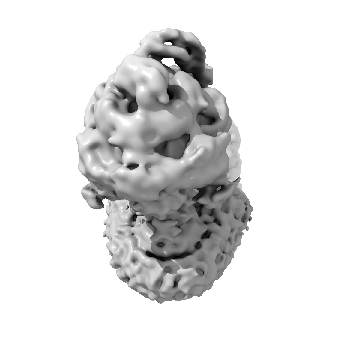
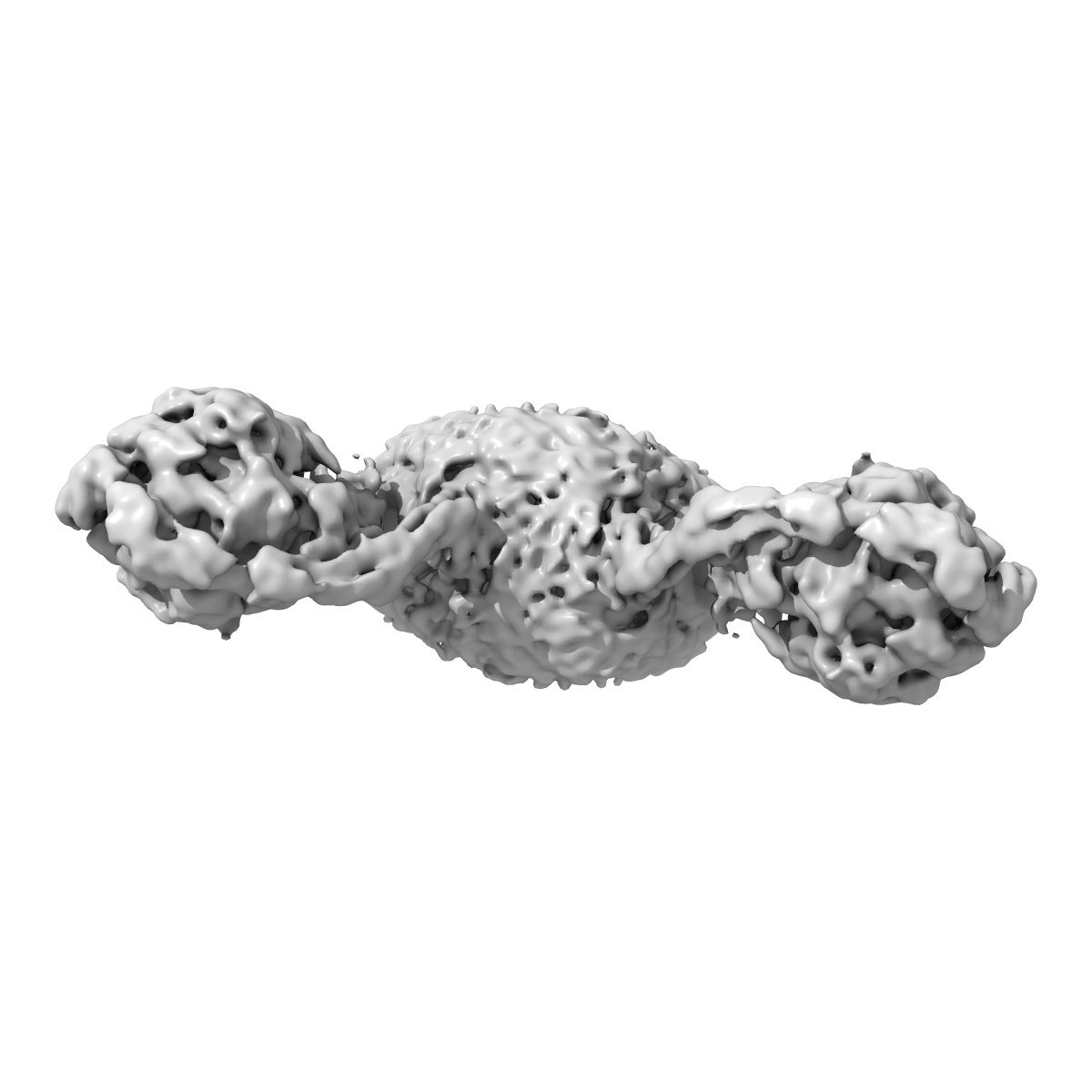
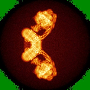
F1Fo ATP synthase dimer from Yarrowia lipolytica
EMD-8151
Single-particle7.7 Å
 Deposition: 20/04/2016
Map released: 20/07/2016
Last modified: 30/08/2017
Sample Organism:
Yarrowia lipolytica
Sample: F1Fo ATP synthase dimer
Deposition Authors: Hahn A, Parey K, Bublitz M, Mills DJ, Zickermann V, Vonck J, Kuehlbrandt W, Meier T
Sample: F1Fo ATP synthase dimer
Deposition Authors: Hahn A, Parey K, Bublitz M, Mills DJ, Zickermann V, Vonck J, Kuehlbrandt W, Meier T
Structure of a Complete ATP Synthase Dimer Reveals the Molecular Basis of Inner Mitochondrial Membrane Morphology.
Hahn A  ,
Parey K ,
Bublitz M ,
Mills DJ ,
Zickermann V,
Vonck J ,
Kuhlbrandt W ,
Meier T
,
Parey K ,
Bublitz M ,
Mills DJ ,
Zickermann V,
Vonck J ,
Kuhlbrandt W ,
Meier T
(2016) Mol. Cell , 63 , 445 - 456
 ,
Parey K ,
Bublitz M ,
Mills DJ ,
Zickermann V,
Vonck J ,
Kuhlbrandt W ,
Meier T
,
Parey K ,
Bublitz M ,
Mills DJ ,
Zickermann V,
Vonck J ,
Kuhlbrandt W ,
Meier T
(2016) Mol. Cell , 63 , 445 - 456
Abstract:
We determined the structure of a complete, dimeric F1Fo-ATP synthase from yeast Yarrowia lipolytica mitochondria by a combination of cryo-EM and X-ray crystallography. The final structure resolves 58 of the 60 dimer subunits. Horizontal helices of subunit a in Fo wrap around the c-ring rotor, and a total of six vertical helices assigned to subunits a, b, f, i, and 8 span the membrane. Subunit 8 (A6L in human) is an evolutionary derivative of the bacterial b subunit. On the lumenal membrane surface, subunit f establishes direct contact between the two monomers. Comparison with a cryo-EM map of the F1Fo monomer identifies subunits e and g at the lateral dimer interface. They do not form dimer contacts but enable dimer formation by inducing a strong membrane curvature of ∼100°. Our structure explains the structural basis of cristae formation in mitochondria, a landmark signature of eukaryotic cell morphology.
We determined the structure of a complete, dimeric F1Fo-ATP synthase from yeast Yarrowia lipolytica mitochondria by a combination of cryo-EM and X-ray crystallography. The final structure resolves 58 of the 60 dimer subunits. Horizontal helices of subunit a in Fo wrap around the c-ring rotor, and a total of six vertical helices assigned to subunits a, b, f, i, and 8 span the membrane. Subunit 8 (A6L in human) is an evolutionary derivative of the bacterial b subunit. On the lumenal membrane surface, subunit f establishes direct contact between the two monomers. Comparison with a cryo-EM map of the F1Fo monomer identifies subunits e and g at the lateral dimer interface. They do not form dimer contacts but enable dimer formation by inducing a strong membrane curvature of ∼100°. Our structure explains the structural basis of cristae formation in mitochondria, a landmark signature of eukaryotic cell morphology.