EMD-8194
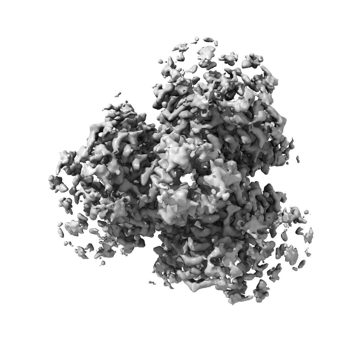
Cryo-EM structure of glutamate dehydrogenase at 1.8 A resolution
EMD-8194
Single-particle1.8 Å
 Deposition: 17/05/2016
Map released: 08/06/2016
Last modified: 06/03/2024
Sample Organism:
Bos taurus
Sample: Glutamate dehydrogenase
Fitted models: 5k12 (Avg. Q-score: 0.598)
Deposition Authors: Merk A, Bartesaghi A
Sample: Glutamate dehydrogenase
Fitted models: 5k12 (Avg. Q-score: 0.598)
Deposition Authors: Merk A, Bartesaghi A
Breaking Cryo-EM Resolution Barriers to Facilitate Drug Discovery.
Merk A,
Bartesaghi A,
Banerjee S,
Falconieri V,
Rao P,
Davis MI  ,
Pragani R,
Boxer MB,
Earl LA,
Milne JL,
Subramaniam S
,
Pragani R,
Boxer MB,
Earl LA,
Milne JL,
Subramaniam S
(2016) Cell , 165 , 1698 - 1707
 ,
Pragani R,
Boxer MB,
Earl LA,
Milne JL,
Subramaniam S
,
Pragani R,
Boxer MB,
Earl LA,
Milne JL,
Subramaniam S
(2016) Cell , 165 , 1698 - 1707
Abstract:
Recent advances in single-particle cryoelecton microscopy (cryo-EM) are enabling generation of numerous near-atomic resolution structures for well-ordered protein complexes with sizes ≥ ∼200 kDa. Whether cryo-EM methods are equally useful for high-resolution structural analysis of smaller, dynamic protein complexes such as those involved in cellular metabolism remains an important question. Here, we present 3.8 Å resolution cryo-EM structures of the cancer target isocitrate dehydrogenase (93 kDa) and identify the nature of conformational changes induced by binding of the allosteric small-molecule inhibitor ML309. We also report 2.8-Å- and 1.8-Å-resolution structures of lactate dehydrogenase (145 kDa) and glutamate dehydrogenase (334 kDa), respectively. With these results, two perceived barriers in single-particle cryo-EM are overcome: (1) crossing 2 Å resolution and (2) obtaining structures of proteins with sizes < 100 kDa, demonstrating that cryo-EM can be used to investigate a broad spectrum of drug-target interactions and dynamic conformational states.
Recent advances in single-particle cryoelecton microscopy (cryo-EM) are enabling generation of numerous near-atomic resolution structures for well-ordered protein complexes with sizes ≥ ∼200 kDa. Whether cryo-EM methods are equally useful for high-resolution structural analysis of smaller, dynamic protein complexes such as those involved in cellular metabolism remains an important question. Here, we present 3.8 Å resolution cryo-EM structures of the cancer target isocitrate dehydrogenase (93 kDa) and identify the nature of conformational changes induced by binding of the allosteric small-molecule inhibitor ML309. We also report 2.8-Å- and 1.8-Å-resolution structures of lactate dehydrogenase (145 kDa) and glutamate dehydrogenase (334 kDa), respectively. With these results, two perceived barriers in single-particle cryo-EM are overcome: (1) crossing 2 Å resolution and (2) obtaining structures of proteins with sizes < 100 kDa, demonstrating that cryo-EM can be used to investigate a broad spectrum of drug-target interactions and dynamic conformational states.