EMD-9328
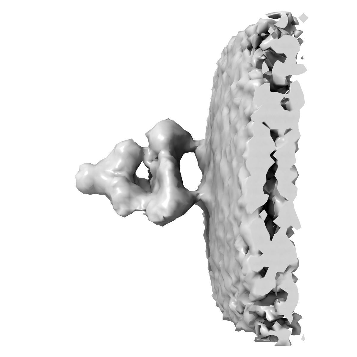
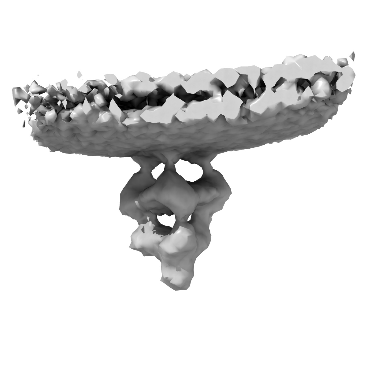
Pre-fusion protein gB revealed on human cytomegalovirus by cryo electron tomography with Volta phase plate
EMD-9328
Subtomogram averaging21.6 Å
 Deposition: 12/11/2018
Map released: 28/11/2018
Last modified: 19/12/2018
Sample Organism:
Human herpesvirus 5 strain AD169
Sample: Human herpesvirus 5 strain AD169
Deposition Authors: Zhu S, Jiayan Z
Sample: Human herpesvirus 5 strain AD169
Deposition Authors: Zhu S, Jiayan Z
Different functional states of fusion protein gB revealed on human cytomegalovirus by cryo electron tomography with Volta phase plate.
Si Z  ,
Zhang J ,
Shivakoti S,
Atanasov I,
Tao CL ,
Hui WH,
Zhou K,
Yu X,
Li W,
Luo M,
Bi GQ ,
Zhou ZH
,
Zhang J ,
Shivakoti S,
Atanasov I,
Tao CL ,
Hui WH,
Zhou K,
Yu X,
Li W,
Luo M,
Bi GQ ,
Zhou ZH
(2018) Plos Pathog , 14 , e1007452 - e1007452
 ,
Zhang J ,
Shivakoti S,
Atanasov I,
Tao CL ,
Hui WH,
Zhou K,
Yu X,
Li W,
Luo M,
Bi GQ ,
Zhou ZH
,
Zhang J ,
Shivakoti S,
Atanasov I,
Tao CL ,
Hui WH,
Zhou K,
Yu X,
Li W,
Luo M,
Bi GQ ,
Zhou ZH
(2018) Plos Pathog , 14 , e1007452 - e1007452
Abstract:
Human cytomegalovirus (HCMV) enters host by glycoprotein B (gB)-mediated membrane fusion upon receptor-binding to gH/gL-related complexes, causing devastating diseases such as birth defects. Although an X-ray crystal structure of the recombinant gB ectodomain at postfusion conformation is available, the structures of prefusion gB and its complex with gH/gL on the viral envelope remain elusive. Here, we demonstrate the utility of cryo electron tomography (cryoET) with energy filtering and the cutting-edge technologies of Volta phase plate (VPP) and direct electron-counting detection to capture metastable prefusion viral fusion proteins and report the structures of glycoproteins in the native environment of HCMV virions. We established the validity of our approach by obtaining cryoET in situ structures of the vesicular stomatitis virus (VSV) glycoprotein G trimer (171 kD) in prefusion and postfusion conformations, which agree with the known crystal structures of purified G trimers in both conformations. The excellent contrast afforded by these technologies has enabled us to identify gB trimers (303kD) in two distinct conformations in HCMV tomograms and obtain their in situ structures at up to 21 Å resolution through subtomographic averaging. The predominant conformation (79%), which we designate as gB prefusion conformation, fashions a globular endodomain and a Christmas tree-shaped ectodomain, while the minority conformation (21%) has a columnar tree-shaped ectodomain that matches the crystal structure of the "postfusion" gB ectodomain. We also observed prefusion gB in complex with an "L"-shaped density attributed to the gH/gL complex. Integration of these structures of HCMV glycoproteins in multiple functional states and oligomeric forms with existing biochemical data and domain organization of other class III viral fusion proteins suggests that gH/gL receptor-binding triggers conformational changes of gB endodomain, which in turn triggers two essential steps to actuate virus-cell membrane fusion: exposure of gB fusion loops and unfurling of gB ectodomain.
Human cytomegalovirus (HCMV) enters host by glycoprotein B (gB)-mediated membrane fusion upon receptor-binding to gH/gL-related complexes, causing devastating diseases such as birth defects. Although an X-ray crystal structure of the recombinant gB ectodomain at postfusion conformation is available, the structures of prefusion gB and its complex with gH/gL on the viral envelope remain elusive. Here, we demonstrate the utility of cryo electron tomography (cryoET) with energy filtering and the cutting-edge technologies of Volta phase plate (VPP) and direct electron-counting detection to capture metastable prefusion viral fusion proteins and report the structures of glycoproteins in the native environment of HCMV virions. We established the validity of our approach by obtaining cryoET in situ structures of the vesicular stomatitis virus (VSV) glycoprotein G trimer (171 kD) in prefusion and postfusion conformations, which agree with the known crystal structures of purified G trimers in both conformations. The excellent contrast afforded by these technologies has enabled us to identify gB trimers (303kD) in two distinct conformations in HCMV tomograms and obtain their in situ structures at up to 21 Å resolution through subtomographic averaging. The predominant conformation (79%), which we designate as gB prefusion conformation, fashions a globular endodomain and a Christmas tree-shaped ectodomain, while the minority conformation (21%) has a columnar tree-shaped ectodomain that matches the crystal structure of the "postfusion" gB ectodomain. We also observed prefusion gB in complex with an "L"-shaped density attributed to the gH/gL complex. Integration of these structures of HCMV glycoproteins in multiple functional states and oligomeric forms with existing biochemical data and domain organization of other class III viral fusion proteins suggests that gH/gL receptor-binding triggers conformational changes of gB endodomain, which in turn triggers two essential steps to actuate virus-cell membrane fusion: exposure of gB fusion loops and unfurling of gB ectodomain.