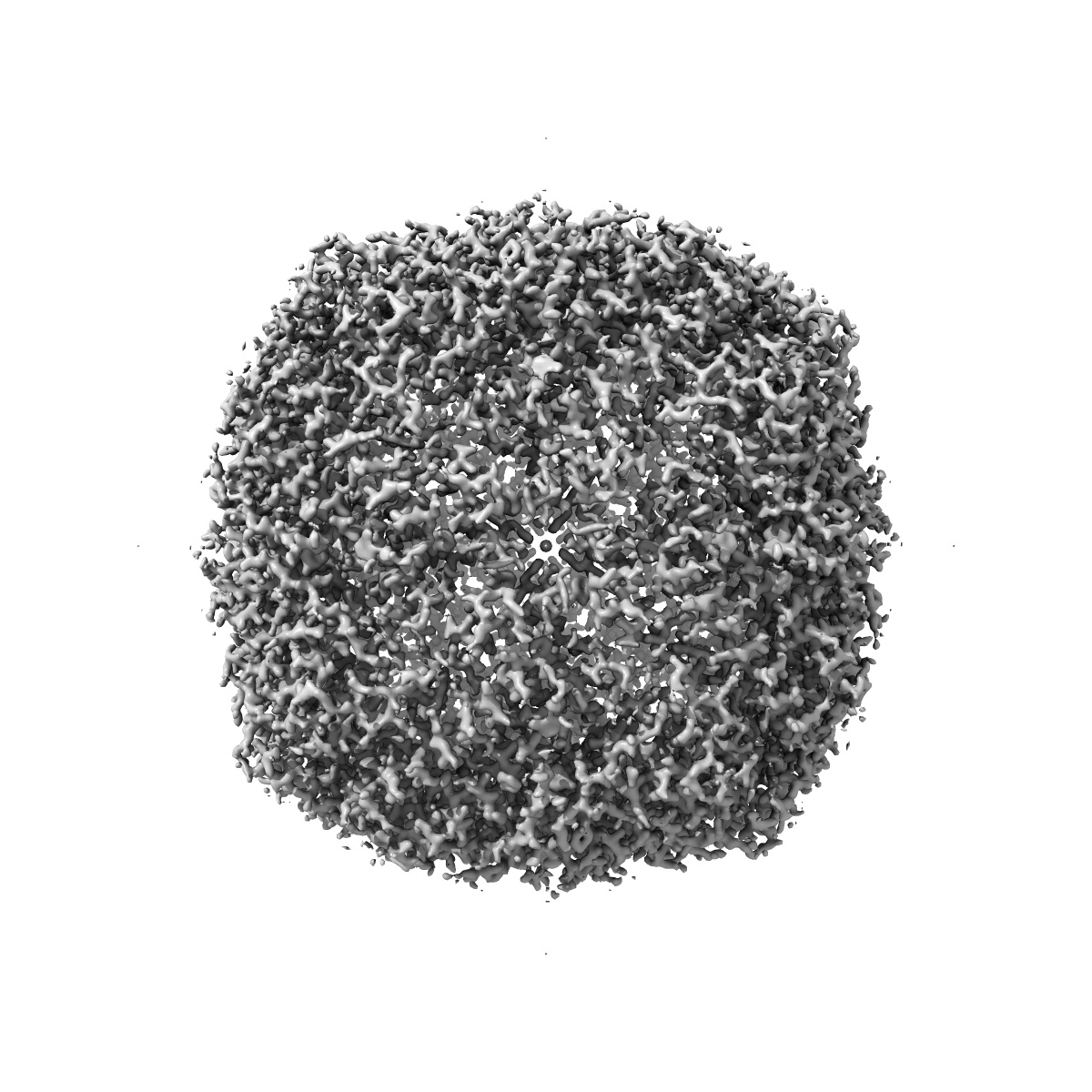
EMD-9890
Cryo-EM structure of human apoferritin at 1.9A resolution by CRYO ARM 300
EMD-9890
Single-particle1.9 Å
 Deposition: 19/04/2019
Map released: 15/05/2019
Last modified: 19/06/2019
Sample Organism:
Homo sapiens
Sample: apoferritin
Deposition Authors: Hamaguchi T, Maki-Yonekura S, Naitow H, Matsuura Y, Ishikawa T, Yonekura K
Sample: apoferritin
Deposition Authors: Hamaguchi T, Maki-Yonekura S, Naitow H, Matsuura Y, Ishikawa T, Yonekura K
A new cryo-EM system for single particle analysis.
Hamaguchi T  ,
Maki-Yonekura S,
Naitow H,
Matsuura Y,
Ishikawa T,
Yonekura K
,
Maki-Yonekura S,
Naitow H,
Matsuura Y,
Ishikawa T,
Yonekura K
(2019) J Struct Biol , 207 , 40 - 48
 ,
Maki-Yonekura S,
Naitow H,
Matsuura Y,
Ishikawa T,
Yonekura K
,
Maki-Yonekura S,
Naitow H,
Matsuura Y,
Ishikawa T,
Yonekura K
(2019) J Struct Biol , 207 , 40 - 48
Abstract:
A new cryo-EM system has been investigated for single particle analysis of protein structures. The system provides parallel illumination of a highly-coherent 300 kV electron beam from a cold-field emission gun, and boosts image contrast with an in-column energy filter and a hole-free phase plate. It includes motorized cryo-sample loading and automated liquid-nitrogen filling for cooling multiple samples. In this study, we describe gun and electron beam characteristics, and demonstrate the suitability of this system for single particle reconstructions. The performance of the system is tested on two examples, a spherical virus and apoferritin. GUI programs have also been developed to control and monitor the system for correct illumination, imaging with less ellipticity and steady magnification, and timing of flashing and liquid-nitrogen filling. These programs are especially useful for efficient application of the system to single particle cryo-EM.
A new cryo-EM system has been investigated for single particle analysis of protein structures. The system provides parallel illumination of a highly-coherent 300 kV electron beam from a cold-field emission gun, and boosts image contrast with an in-column energy filter and a hole-free phase plate. It includes motorized cryo-sample loading and automated liquid-nitrogen filling for cooling multiple samples. In this study, we describe gun and electron beam characteristics, and demonstrate the suitability of this system for single particle reconstructions. The performance of the system is tested on two examples, a spherical virus and apoferritin. GUI programs have also been developed to control and monitor the system for correct illumination, imaging with less ellipticity and steady magnification, and timing of flashing and liquid-nitrogen filling. These programs are especially useful for efficient application of the system to single particle cryo-EM.