EMD-23441
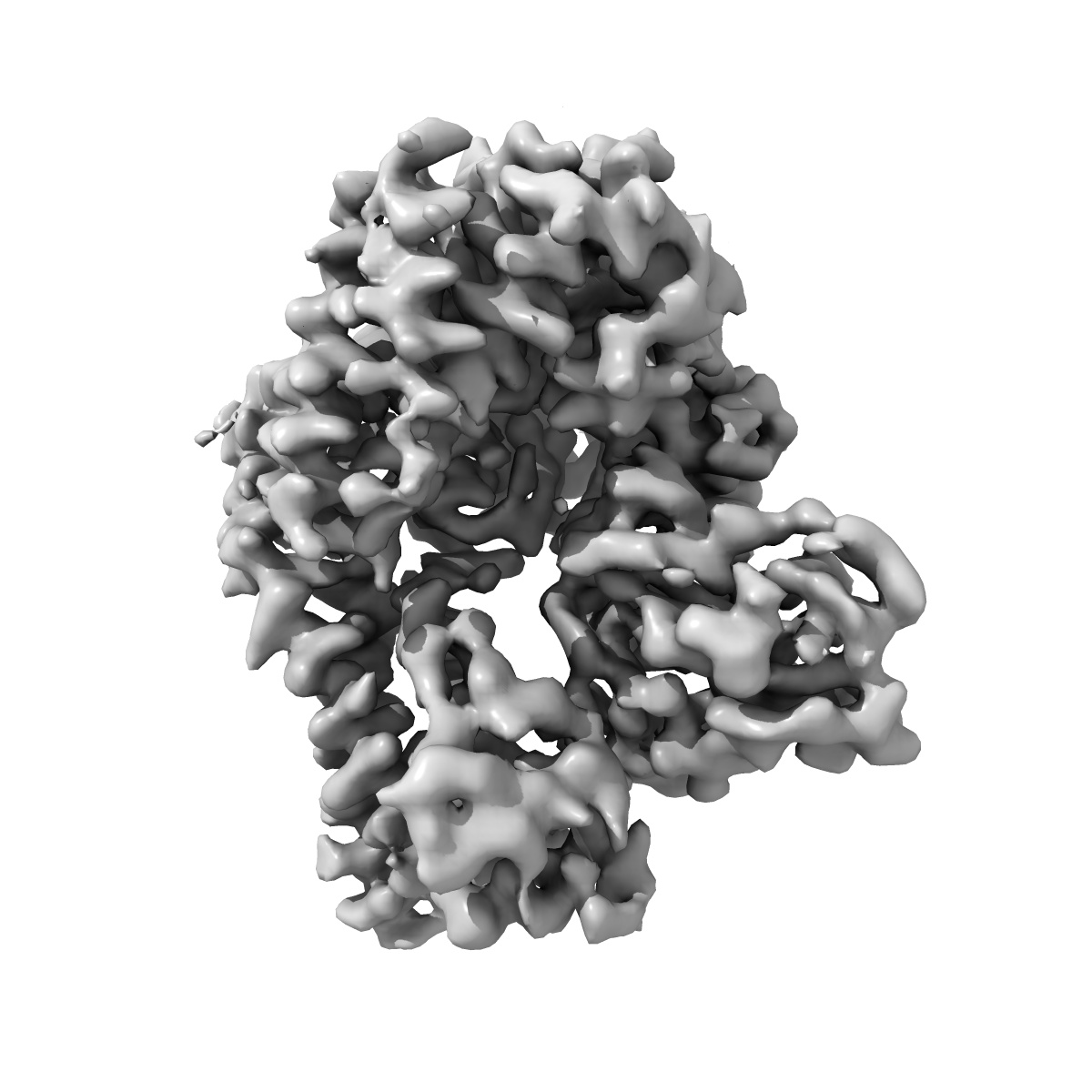
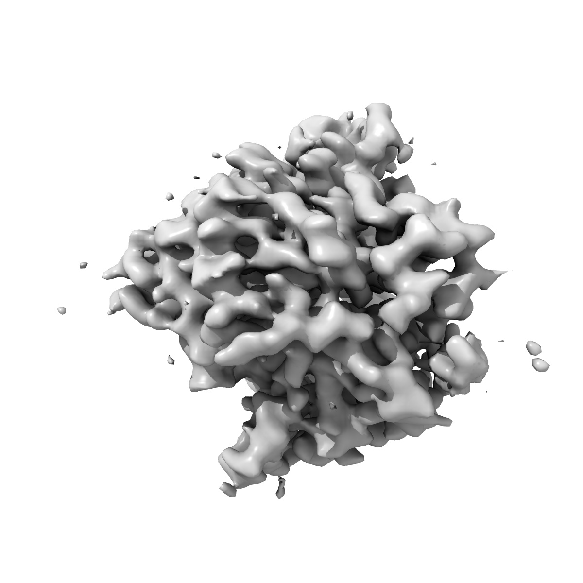
Structure of human SetD3 methyl-transferase in complex with 2A protease from Coxsackievirus B3
EMD-23441
Single-particle3.5 Å
 Deposition: 05/02/2021
Map released: 10/08/2022
Last modified: 29/05/2024
Sample Organism:
Coxsackievirus B3 (strain Nancy),
Coxsackievirus B3 (strain Nancy),
Homo sapiens
Sample: Ternary complex of Coxsackievirus B3 2A protease and human SetD3 methyltransferase
Fitted models: 7lms (Avg. Q-score: 0.452)
Deposition Authors: Verba KA ,
Schulze-Gahmen U
,
Schulze-Gahmen U
Sample: Ternary complex of Coxsackievirus B3 2A protease and human SetD3 methyltransferase
Fitted models: 7lms (Avg. Q-score: 0.452)
Deposition Authors: Verba KA
 ,
Schulze-Gahmen U
,
Schulze-Gahmen U
Structure-function analysis of enterovirus protease 2A in complex with its essential host factor SETD3.
Peters CE ,
Schulze-Gahmen U ,
Eckhardt M ,
Jang GM ,
Xu J,
Pulido EH ,
Bardine C,
Craik CS ,
Ott M ,
Gozani O ,
Verba KA ,
Huttenhain R ,
Carette JE ,
Krogan NJ
(2022) Nat Commun , 13 , 5282 - 5282
,
Schulze-Gahmen U ,
Eckhardt M ,
Jang GM ,
Xu J,
Pulido EH ,
Bardine C,
Craik CS ,
Ott M ,
Gozani O ,
Verba KA ,
Huttenhain R ,
Carette JE ,
Krogan NJ
(2022) Nat Commun , 13 , 5282 - 5282
Abstract:
Enteroviruses cause a number of medically relevant and widespread human diseases with no approved antiviral therapies currently available. Host-directed therapies present an enticing option for this diverse genus of viruses. We have previously identified the actin histidine methyltransferase SETD3 as a critical host factor physically interacting with the viral protease 2A. Here, we report the 3.5 Å cryo-EM structure of SETD3 interacting with coxsackievirus B3 2A at two distinct interfaces, including the substrate-binding surface within the SET domain. Structure-function analysis revealed that mutations of key residues in the SET domain resulted in severely reduced binding to 2A and complete protection from enteroviral infection. Our findings provide insight into the molecular basis of the SETD3-2A interaction and a framework for the rational design of host-directed therapeutics against enteroviruses.
Enteroviruses cause a number of medically relevant and widespread human diseases with no approved antiviral therapies currently available. Host-directed therapies present an enticing option for this diverse genus of viruses. We have previously identified the actin histidine methyltransferase SETD3 as a critical host factor physically interacting with the viral protease 2A. Here, we report the 3.5 Å cryo-EM structure of SETD3 interacting with coxsackievirus B3 2A at two distinct interfaces, including the substrate-binding surface within the SET domain. Structure-function analysis revealed that mutations of key residues in the SET domain resulted in severely reduced binding to 2A and complete protection from enteroviral infection. Our findings provide insight into the molecular basis of the SETD3-2A interaction and a framework for the rational design of host-directed therapeutics against enteroviruses.