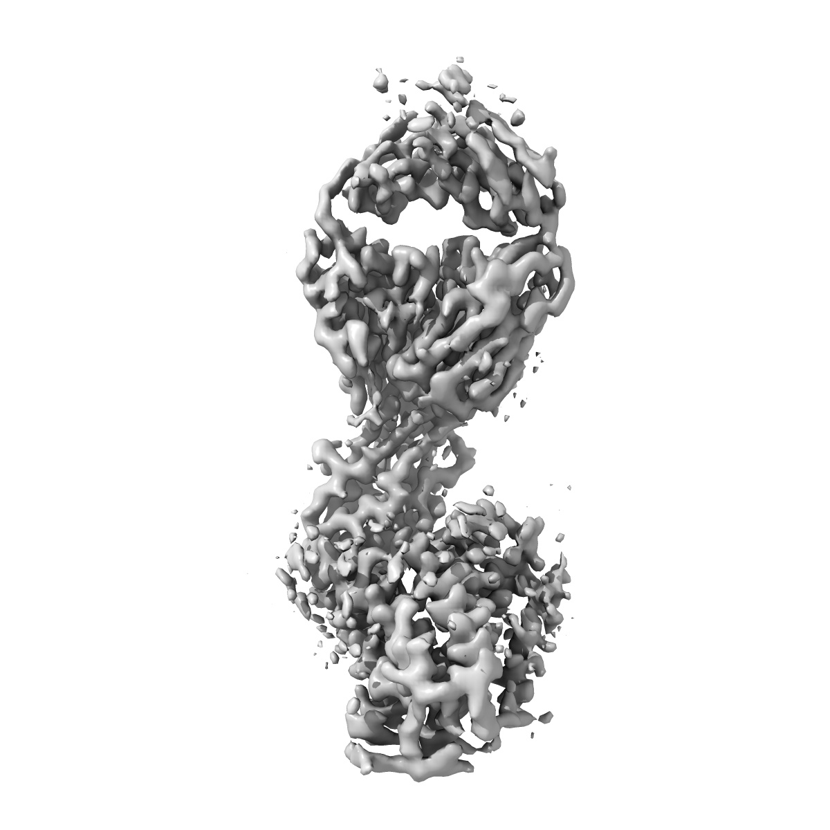
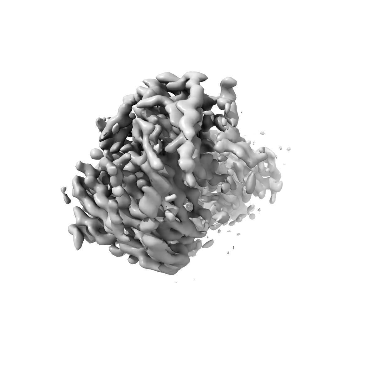
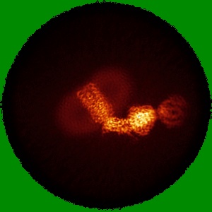
EMD-27489
Cryo-EM structure of cystinosin in a lumen-open state
EMD-27489
Single-particle3.32 Å
 Deposition: 05/07/2022
Map released: 21/09/2022
Last modified: 23/10/2024
Sample Organism:
Mus musculus,
Homo sapiens
Sample: Cystinosin-Fab 3H5 complex in lumen-open conformation
Fitted models: 8dki (Avg. Q-score: 0.497)
Deposition Authors: Schmiege P ,
Li X
,
Li X
Sample: Cystinosin-Fab 3H5 complex in lumen-open conformation
Fitted models: 8dki (Avg. Q-score: 0.497)
Deposition Authors: Schmiege P
 ,
Li X
,
Li X
Structure and mechanism of human cystine exporter cystinosin.
Guo X,
Schmiege P ,
Assafa TE ,
Wang R ,
Xu Y,
Donnelly L,
Fine M,
Ni X,
Jiang J,
Millhauser G,
Feng L,
Li X
(2022) Cell , 185 , 3739 - 3752.e18
,
Assafa TE ,
Wang R ,
Xu Y,
Donnelly L,
Fine M,
Ni X,
Jiang J,
Millhauser G,
Feng L,
Li X
(2022) Cell , 185 , 3739 - 3752.e18
Abstract:
Lysosomal amino acid efflux by proton-driven transporters is essential for lysosomal homeostasis, amino acid recycling, mTOR signaling, and maintaining lysosomal pH. To unravel the mechanisms of these transporters, we focus on cystinosin, a prototypical lysosomal amino acid transporter that exports cystine to the cytosol, where its reduction to cysteine supplies this limiting amino acid for diverse fundamental processes and controlling nutrient adaptation. Cystinosin mutations cause cystinosis, a devastating lysosomal storage disease. Here, we present structures of human cystinosin in lumen-open, cytosol-open, and cystine-bound states, which uncover the cystine recognition mechanism and capture the key conformational states of the transport cycle. Our structures, along with functional studies and double electron-electron resonance spectroscopic investigations, reveal the molecular basis for the transporter's conformational transitions and protonation switch, show conformation-dependent Ragulator-Rag complex engagement, and demonstrate an unexpected activation mechanism. These findings provide molecular insights into lysosomal amino acid efflux and a potential therapeutic strategy.
Lysosomal amino acid efflux by proton-driven transporters is essential for lysosomal homeostasis, amino acid recycling, mTOR signaling, and maintaining lysosomal pH. To unravel the mechanisms of these transporters, we focus on cystinosin, a prototypical lysosomal amino acid transporter that exports cystine to the cytosol, where its reduction to cysteine supplies this limiting amino acid for diverse fundamental processes and controlling nutrient adaptation. Cystinosin mutations cause cystinosis, a devastating lysosomal storage disease. Here, we present structures of human cystinosin in lumen-open, cytosol-open, and cystine-bound states, which uncover the cystine recognition mechanism and capture the key conformational states of the transport cycle. Our structures, along with functional studies and double electron-electron resonance spectroscopic investigations, reveal the molecular basis for the transporter's conformational transitions and protonation switch, show conformation-dependent Ragulator-Rag complex engagement, and demonstrate an unexpected activation mechanism. These findings provide molecular insights into lysosomal amino acid efflux and a potential therapeutic strategy.