EMD-36861
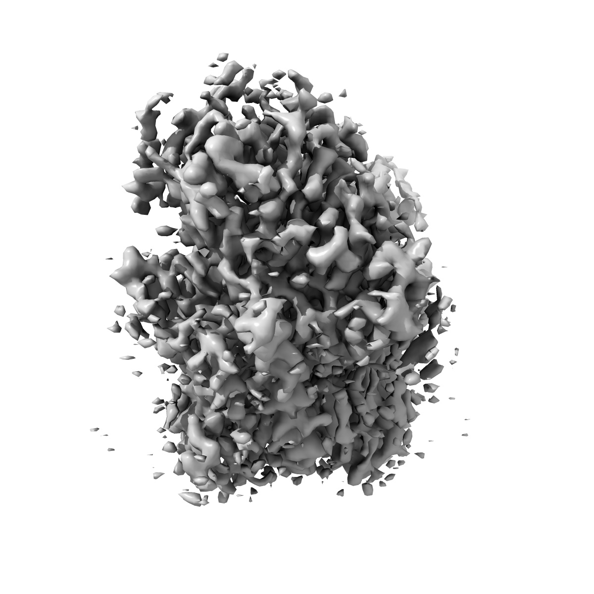
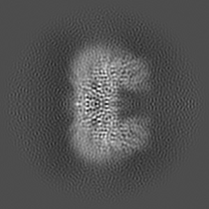
S. cerevisiae Chs1 in complex with UDP and GlcNAc
EMD-36861
Single-particle3.06 Å
 Deposition: 17/07/2023
Map released: 06/09/2023
Last modified: 08/05/2024
Sample Organism:
Saccharomyces cerevisiae
Sample: S. cerevisiae Chs1 in complex with UDP and GlcNAc
Fitted models: 8k3u (Avg. Q-score: 0.445)
Deposition Authors: Bai L ,
Chen D
,
Chen D
Sample: S. cerevisiae Chs1 in complex with UDP and GlcNAc
Fitted models: 8k3u (Avg. Q-score: 0.445)
Deposition Authors: Bai L
 ,
Chen D
,
Chen D
Structure, catalysis, chitin transport, and selective inhibition of chitin synthase.
Abstract:
Chitin is one of the most abundant natural biopolymers and serves as a critical structural component of extracellular matrices, including fungal cell walls and insect exoskeletons. As a linear polymer of β-(1,4)-linked N-acetylglucosamine, chitin is synthesized by chitin synthases, which are recognized as targets for antifungal and anti-insect drugs. In this study, we determine seven different cryo-electron microscopy structures of a Saccharomyces cerevisiae chitin synthase in the absence and presence of glycosyl donor, acceptor, product, or peptidyl nucleoside inhibitors. Combined with functional analyses, these structures show how the donor and acceptor substrates bind in the active site, how substrate hydrolysis drives self-priming, how a chitin-conducting transmembrane channel opens, and how peptidyl nucleoside inhibitors inhibit chitin synthase. Our work provides a structural basis for understanding the function and inhibition of chitin synthase.
Chitin is one of the most abundant natural biopolymers and serves as a critical structural component of extracellular matrices, including fungal cell walls and insect exoskeletons. As a linear polymer of β-(1,4)-linked N-acetylglucosamine, chitin is synthesized by chitin synthases, which are recognized as targets for antifungal and anti-insect drugs. In this study, we determine seven different cryo-electron microscopy structures of a Saccharomyces cerevisiae chitin synthase in the absence and presence of glycosyl donor, acceptor, product, or peptidyl nucleoside inhibitors. Combined with functional analyses, these structures show how the donor and acceptor substrates bind in the active site, how substrate hydrolysis drives self-priming, how a chitin-conducting transmembrane channel opens, and how peptidyl nucleoside inhibitors inhibit chitin synthase. Our work provides a structural basis for understanding the function and inhibition of chitin synthase.